


A timely referral means a happy patient out of pain
Featured Products Promotional FeaturesPosted by: Dental Design 14th April 2021
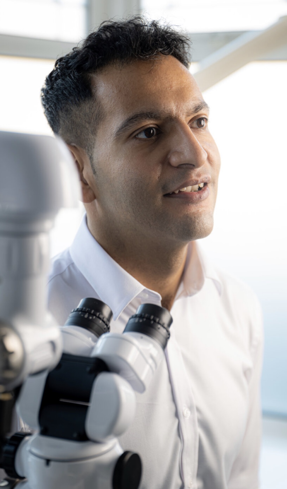
Dr Dhiraj Arora qualified in 2007 from Kings College London and, after five years as an associate dentist, is now the owner of two practices. Having completed his Masters in Endodontic Practice from QMUL, he now principally works as an endodontist across multiple practices in London.
Dhiraj has been involved in the mentorship of newly qualified dentists and is an educational supervisor within the London Deanery. This, along with being an Honorary Lecturer in Endodontics (QMUL) allows him to combine his enthusiasm for endo and teaching. He has recently set up Evo Endo to provide postgraduate endodontic courses to general dental practitioners.
Initial presentation and background
Visit 1:
The patient was a referral for root canal treatment (RCT) on LL6. She was triaged for COVID-19 over the telephone and also on entering the surgery. For the appointment, I wore the following PPE: respirator mask, gown, face shield, loupes and gloves.
The patient said she had been advised by her general dentist that she required RCT. She had been in continuous pain after she’d had a crown fitted and the GDP did try to find the nerves; however, this did not help. The patient told me that the tooth hadn’t felt right since the crown was done and that she would like the pain to stop. She had been taking painkillers and prescribed a course of antibiotics.
Results of examinations
Extraorally, no abnormality was detected. Intraoral exam on LL6 revealed a recent onlay had been carried out and attempted extirpation, but only one canal had been located.
Mobility was grade 0. The tooth was slightly tender to percussion, also there was mild buccal tenderness to palpation. There was no endo-related pocketing when the probe was walked around the gingival crevice. No evidence of a sinus tract or intraoral swelling were found.
A pre-operative radiograph showed the onlay in close association to the pulp space, with no obvious pathosis (see Figure 1).
Diagnosis
A diagnosis was made for the LL6 of symptomatic apical periodontitis with previously initiated treatment.
The options regarding this tooth were:
1) Do nothing. However, the patient was warned that, if left untreated, it could cause an acute flare up at any time and have a reduced prognosis due to persistent infection.
2) Root canal treatment (RCT) with cuspal coverage.
3) Extraction.
The patient was happy to proceed with RCT. Consent was discussed, with the risks and benefits explained, including the risk of re-infection and the complex anatomy. The consent form was duly signed, witnessed by me and the dental nurse.
Treatment pathway
Local anaesthesia was administered: 4.4 ml lidocaine hydrochloride, 2% 1:80,000 adrenaline via buccal infiltration and inferior dental block (IDB).
A rubber dam was placed over LL6, with a clamp and rubber dam liquid used to provide a tight seal. The tooth was accessed (see Figure 2), with three orifices located and checks made for a potential second distal canal. No second distal canal was found, with a note made to check again at the next visit. The located distal canal was oval-shaped and centrally located.
A glidepath was created using K-Flex Files 06, 08, 10 and 15. Throughout the procedure, the canal was irrigated with an enhanced sodium hypochlorite solution applied from a syringe.
Working lengths were established using an apex locator: Mesiobuccal (MB), 18 mm (reference point, buccal cavity wall); Mesiolingual (ML), 18 mm (reference point, mesiolingual cusp) and Distal (D), 19.5 mm (reference point, mesiolingual cusp).
Apical preparation was completed using COLTENE’s HyFlex™ EDM files and CanalPro™ Jeni motor sequentially: the 10/.05 Glidepath file then the 25/~ OneFile. These files definitely know their way around curves. Due to their controlled memory, the files follow the anatomy of the canal, thus significantly reducing the risk of ledging, transportation or perforation of a canal. Like stainless steel files, HyFlex™ files can be pre-bent. Used in combination with the CanalPro™ Jeni, which uses complex algorithms to control file movement at millisecond intervals, I am able to navigate the canal system to achieve safe, effective and predictable mechanical preparations for our patients. The motor is smooth and efficient, and the audible signals provides the clinician with a regular reminder of the importance of constant irrigation. The Jeni has an integrated apex locator, useful as it provides information regarding the positioning of rotary files within the canals at all times, which could potentially reduce the incidence of overpreparation.
The canals were prepared to the above sizes, with continuous irrigation using the sodium hypochlorite solution. The canals were dried with paper points, a calcium hydroxide paste placed in them and a pledget was used in the pulp chamber space. The tooth was temporarily restored, with occlusion and contacts checked.
The patient was told to expect some post-op pain and tenderness and advised to take painkillers and avoid having anything hard on the tooth, due to the risk of fracture. She was also advised about the possibility of an acute flare-up and/or swelling in the area, alongside the possibility of tenderness from the jaw joint. If any of these scenarios were to occur, she was to use anti-inflammatories, cold compresses and contact the dental practice. The patient understood these instructions and left happy.
Visit 2:
For her next visit, the plan was to obturate and provide a coronal seal with a direct permanent restoration. Once again, the patient was triaged for COVID-19 over the telephone and on entering the surgery. I wore a respirator mask, gown, face shield, loupes and gloves.
The patient was able to report that since the last visit she’d experienced no pain or discomfort, so would like the treatment completed. She was made aware of the risks and confirmed consent, as before.
I was then able to proceed with the continuation of the RCT on LL6. Once again, it was delivered under local anaesthesia: 4.4ml lidocaine hydrochloride, 2% 1:80,000 adrenaline via buccal infiltration and IDB.
A rubber dam was placed over LL6, with a clamp and rubber dam liquid used to provide a tight seal. The tooth was accessed, three orifices relocated and rechecked for the presence of a potential second distal canal, using a microscope and ultrasonics.
Working lengths in each canal were re-established and apical preparation completed using COLTENE’s HyFlex™ EDM file system. Apical gauging was carried out to determine the size of the finishing files required: MB, binding file ISO size 25, finishing file 25/~ OneFile; ML, binding file ISO size 25, finishing file 25/~ OneFile and D, binding file ISO size 25, finishing file 25/~ OneFile.
The canals were prepared to the above sizes, with continuous irrigation throughout with sodium hypochlorite solution applied from a syringe.
Matching Gutta-Percha points were placed in situ (see Figure 3) and a mastercone long cone periapical radiograph (LCPA) of the LL6 was performed (see Figure 4), to assess the apical extent of the master gutta percha cones. Reporting showed a potential obturation to within 2 mm of radiographic apex.
I performed mechanical agitation of the irrigant, with well-fitting gutta percha cones using long vertical strokes. I then gave a penultimate rinse with EDTA 17% for 1 minute, followed by the sodium hypochlorite. The canals were dried with paper points.
The canals were obturated using a continuous wave of condensation (warm vertical) technique: matching master cones with sealant to working length. A heat source was used to remove the coronal portion of the cones, to a binding point 5 mm short of the working length. The canals were backfilled with thermoplastic gutta percha (see Figures 5 and 6).
The tooth was etched, bonded and restored with SDR as an orifice seal/base and COLTENE’s EverGlow™ universal composite. Occlusion and contacts were checked.
The patient was advised to take painkillers to relieve any discomfort and to expect some pain and possible tenderness of the tooth. As per visit one, she was also advised about the possibility of an acute flare-up and/or swelling in the area, or the possibility of tenderness from the jaw joint. If any of these scenarios occur, she was to use anti-inflammatories, cold compresses and contact the dental practice. The patient understood these instructions.
A LCPA radiograph was taken, justified for post-operative assessment of endodontic treatment and to check that a coronal seal was provided (see Figure 7). Reporting showed the RCT well condensed and within 2mm of the radiographic apex. The patient was advised to see her GDP for review and left happy (see Figure 8).
Images


For more on COLTENE, visit: www.coltene.com,
email info.uk@coltene.com or call 01444 235486.
For endodontic courses, visit: www.evoendo.co.uk
email enquiries@evoendo.co.uk follow drdij_evoendo on Instagram







