


Periodontal surgical treatment for recession
Featured Products Promotional FeaturesPosted by: Dental Design 17th February 2023
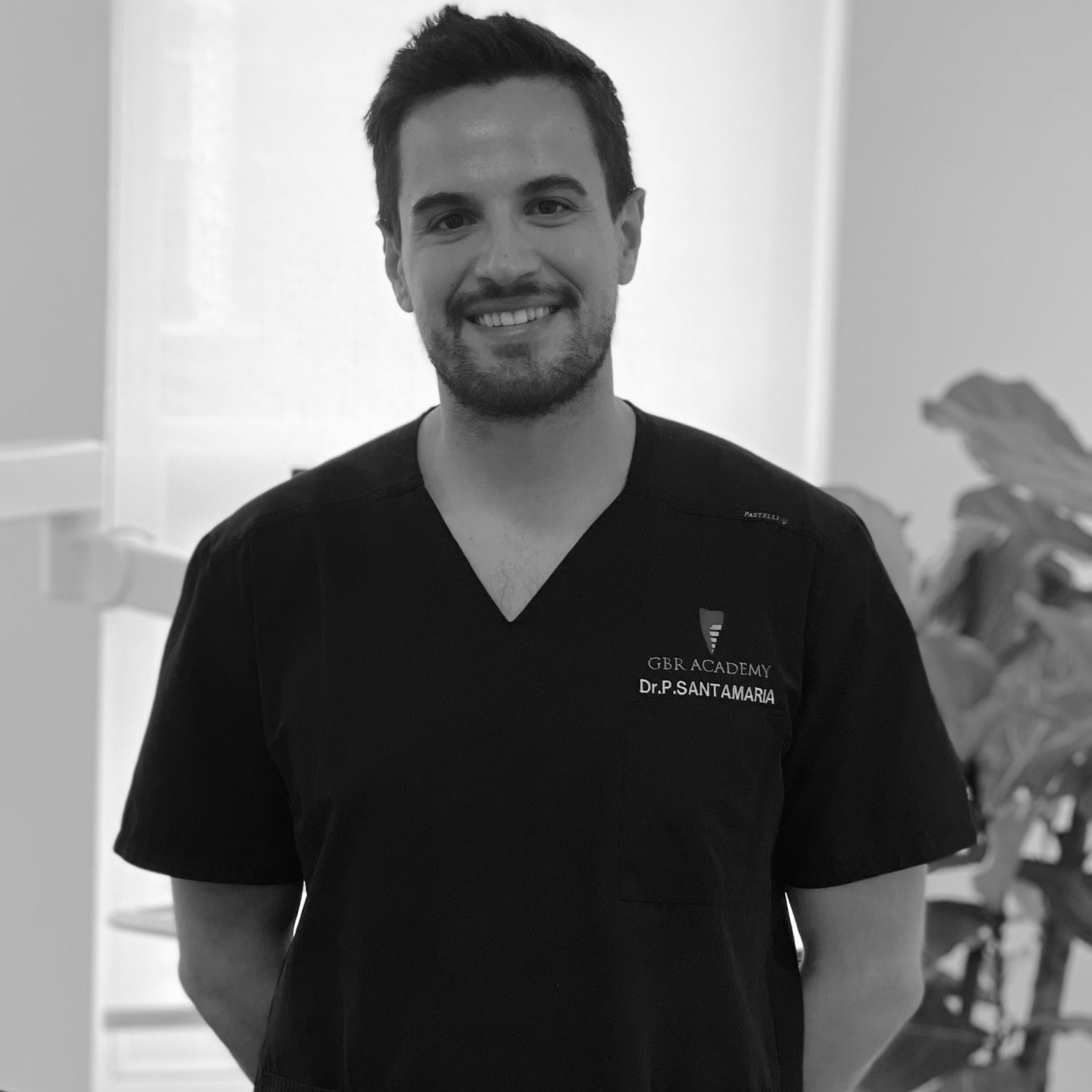
Dr Pasquale Santamaria, specialist oral surgeon and expert in periodontology at Pond Square Dental in London, presents a case he recently completed. This patient presented with gingival recession following orthodontic treatment. They reported an aesthetic issue related to the LR2 and high sensitivity.
Assessment
A full periodontal assessment was conducted, including evaluation of pocket depth, severity of gingival recession, gingival phenotype analysis, plaque and bleeding score and radiographic analysis of the bone level. A diagnosis was made of 5mm gingival recession on the LR2, with no interdental attachment loss. It was a class 2 (Miller), RT1 (Cairo) as the patient had a thin scalloped gingival phenotype associated with tooth sensitivity and aesthetic issues.
All the available treatment options were discussed in detail with the patient. The two potential solutions in this case were different plastic periodontal surgical techniques:
- Raise a flap and place an autogenous connective tissue graft.
- Raise a flap and place an acellular collagen matrix.
Treatment planning
According to evidence-based medicine, the autogenous graft demonstrates a predictable result in increasing the gingival phenotype. It was selected for this case in agreement with the patient.
Gingival phenotype refers to the three-dimensional gingival volume covering the buccal bone plate; it may be affected by environmental factors and modified by clinical interventions. Gingival phenotype is determined by gingival thickness and keratinised tissue width. These anatomic features allow us to classify the “phenotypes” into three groups: thin scalloped, thick flat and thick scalloped.
It is commonly accepted that thin phenotypes are more prone to developing gingival recessions; similarly, growing gingival recession lesions are often associated with thin phenotypes. Therefore, one of the goals of periodontal plastic surgery should be to obtain a thick gingival unit, in turn reducing the risk of future gingival recession recurrence – especially when the initial flap thickness is less than 0.8mm. For this reason, the additional use of a connective tissue graft collected from the palate seems to be an effective solution in improving gingival phenotype, producing a more predictable result in the long-term. However, this procedure results in a second surgical wound on the palate and longer operative times. Furthermore, high post-operative pain levels can be experienced by patients undergoing this procedure.
For this particular case, it was decided to proceed with a flap and autogenous connective tissue graft. Treatment was planned in detail using digital intraoral scans and digital radiographs. The treatment, postoperative healing and complications, and potential outcomes were discussed with the patient, who provided informed consent to proceed.
Treatment
The procedure began with root debridement, which was accurately performed using sharp curettes. First, two horizontal incisions were performed mesial and distal to the gingival recession, at a distance from the tip of the anatomical papillae equal to the depth of the recession plus 1mm. The achieved flap was elevated using a split-full-split approach until the cementoenamel junction (CEJ) of the tooth could be passively covered by the marginal portion of the flap.
The anatomic papillae coronal to the horizontal incisions were de-epithelised to supply a connective tissue surface to which the flap’s surgical papillae were sutured. The graft was positioned to cover the CEJ, stabilised by sutures and covered by the flap that was positioned leaving the graft exposed.
The connective tissue graft was harvested from the palatal area with two incisions delimitating the donor area. The graft was separated from the underlying tissues with the scalpel’s blade oriented parallel to the palatal surface to obtain a graft thickness of about 2mm. Then, the graft was de-epithelialised by a 15c blade and the fatty tissue was eliminated until a graft with a thickness of about 1.5mm was obtained.
Outcome
At the two-week review, residual stiches were removed – the graft was still healing but looked promising. At the 12-month review, the healing process was complete and the gingival margin covered the CEJ, offering the patient a complete root coverage, no sensitivity, a thick gingival phenotype and an optimal aesthetic outcome.
Discussion
The most important step to approach a patient showing gum recession is the correct diagnosis: including both aetiology and gingival parameters (recessions, thickness and amount of residual keratinised tissues, root convexity and tooth positions). The choice of the surgical techniques should meet the patient’s expectations for both aesthetic and/or functional results. It’s always indicated to have an extensive discussion with the patient, highlighting all risks and potential final outcomes after the procedure, especially in patients with high aesthetic needs.

Figure 1 – Gingival recession upon presentation

Figure 2 – Flap raised less than 0.8mm in thickness

Figure 3 – Graft harvested from palate

Figure 4 – Autogenous connective tissue graft

Figure 5 – Graft placed and sutured in place

Figure 6 – Review appointment 2 weeks post surgery

Figure 7 – 12 months post-operative
For more information on services available and to refer your patient to Dr Santamaria, or to the several other specialists working at Pond Square Dental, please visit www.pondsquaredental.co.uk
Author bio:
Dr Santamaria qualified in Italy in 2015, before undertaking a 24-month postgraduate course in Periodontology and working as teaching assistant from 2016 to 2021 in the Periodontal Unit of the G.D’annunzio University in Italy, focusing on the microbiology aspect of the gum disease.
Dr Santamaria is actively involved in international research in Periodontology. In Italy, he was awarded a research fellowship for two years to look into the role of stem cells in bone regeneration. In 2020, he has completed an annual International Master’s in Research in Dentistry at the UIC University in Barcelona.
Dr Santamaria is the coordinator of an international research project sponsored by Guy’s and St Thomas Hospital in London, focused on the survival of teeth affected by advanced gum disease. He is a PhD candidate in Periodontology at the King’s College London.
Dr Santamaria is also a registered Specialist in Oral Surgery in the UK, after completing a Master’s degree. He is a member of the British Society of Periodontology (BSP), the Furcation European Research Group (FERG), and the Italian Society of Periodontology (SIDP)







