


An anterior aesthetic fixed ortho case
Featured Products Promotional FeaturesPosted by: Dental Design 20th September 2023
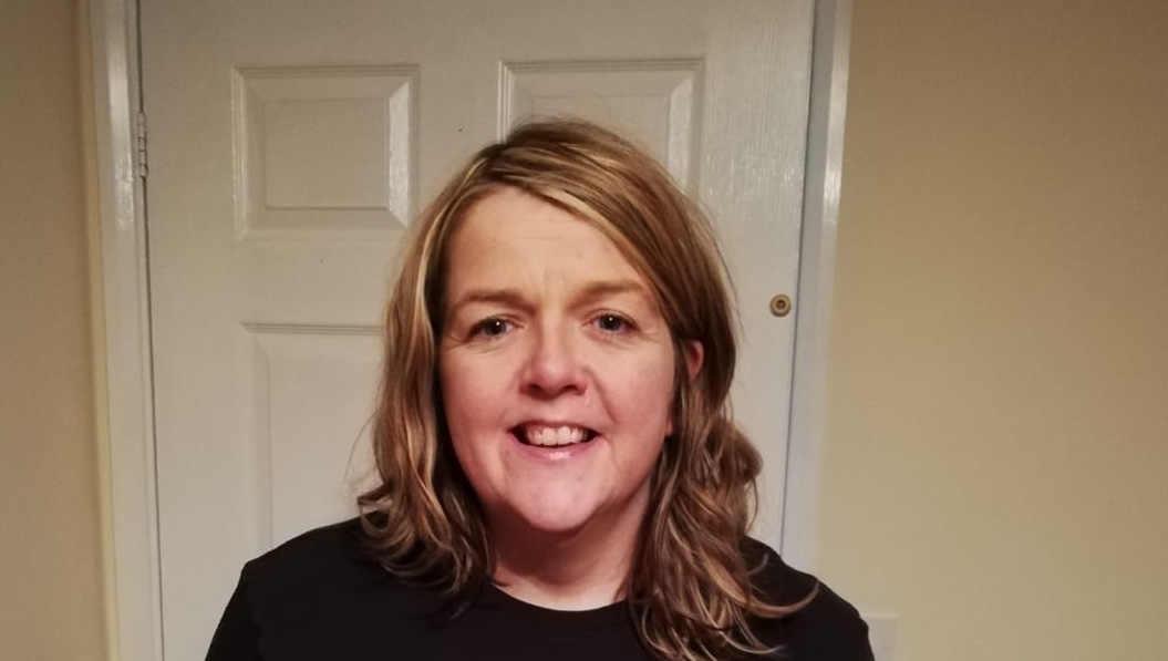
Patient presentation
A 55-year-old female patient was referred internally as she was unhappy with the position of her upper anterior teeth – in particular the relationship between UR1 and UL1. She was also unhappy with the position of her lower teeth but, for financial reasons, did not want to intervene here at this time. Her assessment appointment was in July 2022 and she was keen for treatment to be completed before her wedding in September 2023.
Orthodontic and clinical assessment
A full examination was carried out. The patient presented with upper mild and lower moderate crowding in the anterior segments on a mild class II skeletal base (fig. 1-4). There were no carious lesions, her gingiva was healthy, and radiographs indicated that her bone levels, root morphology, and associated bone were good. The FMPA and lower facial height were average. She had a class I incisal relationship, class I canines and class II molars. There were no crossbites, facial asymmetry or displacements on closure, and the overbite was average. The centrelines were non-coincident with the upper deviated to the right by 1mm. Her periodontal condition was good and her oral hygiene was excellent. The U4’s had already been extracted, and the LR6 was also missing and had been replaced by an implant.
Treatment options
After completing the clinical assessment, the following treatment options were presented to the patient:
- Do nothing
- Referral to a specialist orthodontist
- Removable aligners (SureSmile)
- Fixed appliances (IAS Fixed Braces)
- Composite bonding without orthodontics
- Ceramic veneers
The patient was provided with a mock up regarding composite bonding without straightening first, but she decided against this option. Ceramic veneers would have been too invasive. She preferred the case to be carried out in-house, rather than being seen by a specialist orthodontist. Her only concerns were her upper “social six” so she opted for anterior aesthetic orthodontics (AAO).
We discussed the pros and cons of both fixed and removable solutions, including accepting the already present lower crowding, the centreline deviation and the class II molars. She opted for the fixed option as she knew she would not be able to wear clear aligners for 22 hours a day. Post orthodontic composite bonding was discussed, but the patient was not interested in whitening her teeth.
Treatment planning
The patient’s scans and photographs were sent to the IAS lab. An Archwize™ simulation of the final results was produced, and the patient provided informed consent.
After a face-to-face discussion with the patient, a report was sent to her which included all possible pros and cons of orthodontic treatment including all those not already mentioned. It stressed what would be required of the patient, including appliance care, good oral hygiene, and attending regular appointments. Also included was a breakdown of costs and an estimated treatment timeline, including the post-orthodontic composite bonding.
Treatment provision
Once the signed report was returned, the lab work was ordered from the IAS lab. This included pre-loaded indirect bonding trays for the brackets, thus removing the stress of trying to position them correctly. The patient attended for her fit appointment in August 2022, where all required interproximal reduction (IPR) and progressive proximal reduction (PPR) was carried out. A 0.012 NiTi wire was tied in. She returned regularly over the course of 12 weeks where a 0.016 NiTi wire was placed, the bracket on UL1 was positioned more mesially to help de-rotate the tooth, and a 0.020 x 0.020 was tied.
She returned 1 week later as she was concerned that her teeth were not moving correctly. On examination all was ok, and the patient was reassured. At her review appointment, I decided to reposition the UL1 bracket again more mesially as its position wasn’t quite right. I retied a 0.016 wire to fully ligate the UL1 bracket. Following this, the patient returned requesting removal of the brace as she was very happy with the result. On examination, even though the position of UL1 was much improved, I felt it could be better. The patient stated that she was happy as was, and was scanned for a fixed retainer.
When the patient returned for fixed retainer fitting, and to be scanned for the removable retainer, she had changed her mind and wanted the UR1 and UL1 to be more aligned. In her previous appointment, the patient had signed a waiver, which confirmed that she had requested removal of the brace against my advice. Because of this, she would need to cover the cost for anything else that may involve more lab work. However, I wanted to deliver a good outcome, so I took photos and posted them on the IAS forum to find another solution. As per their suggestion, a little more IPR was carried out and the 0.020 x 0.020 NiTi wire was re-tied. In this instance, no extra charge was applied as the brackets were still fixed. The patient returned 2 weeks later and she stated that she was happy with the results (fig. 5-7).
Not much had changed in this time, but the patient was desperate for removal. She signed a second waiver. The fixed brace was removed after fitting the previously ordered fixed retainer, as the position of the teeth had not changed (fig. 8-9). She was scanned for a removable retainer.
Following this, she emailed me to say that she had made a big mistake, and that she would like to carry on with treatment. The patient attended the practice the same day, and updated scans and photos were posted on the IAS forum. I stressed again that she would need to cover the cost for extra treatment, as her original fixed brace had now been removed.
The best option was to carry out an Archwize™ simulation, for a refiner aligner, requiring the removal of the fixed retainer. This was carried out after a removable retainer was provided, to prevent any relapse. The patient was shown the Archwize™ 3D simulation after refiners, and was happy with the projected outcome. The treatment plan required 3 refiners to be worn 22 hours per day, and changed every 2 weeks. After treatment, the patient was happy with the results.
Composite bonding was then carried out to close the black triangles between UR1 and UL1, and UL1 and UL2, as well as along the edge of UL2. The patient was then scanned for a fixed retainer which was fitted a week later.
The patient’s treatment was completed 4 months before her wedding! (fig. 10-12)
Case reflection
There were definitely a couple of lessons learned whilst doing this case.
Firstly, if you are not happy with your patients’ decisions, advise them to carry on with their treatment. Failing that, ensure that the fact that they are acting against your advice is recorded in writing. It can get expensive for you otherwise.
Secondly, use a good camera for high-quality dental photographs!

Fig 1. Initial Presentation

Fig 2. Initial Presentation

Fig 3. Initial Presentation

Fig 4. Initial Presentation

Fig 5. Repositoned bracket

Fig 6. Repositoned bracket

Fig 7. Repositoned bracket

Fig 8. Fixed retainer

Fig 9. Fixed retainer

Fig 10. Finished treatment

Fig 11. Finished treatment

Fig 12. Finished treatment
For more information on upcoming IAS Academy training courses, please visit
www.iasortho.com or call 01932 336470 (Press 1)
Author Bio:
Dr Sarah Canavan has extensive dental experience dating back to 1995 with qualifications including a master’s degree in Pain Management and a Postgraduate Certificate in Contemporary Restorative and Aesthetic Dentistry. She provides orthodontic treatment using fixed braces, Quick Straight Teeth, and other aligner systems, in addition to providing composite bonding.







