


Paying attention to dimension
Featured Products Promotional FeaturesPosted by: Dental Design 22nd November 2023
Exploring the advances in dental radiography
Across the UK, more dental radiographs are taken each year than any other form of x-ray diagnostic image. The various forms of dental x-ray imaging now available have become indispensable tools in the efficient diagnosis of disease and the effective planning of treatment.[i]
Dental radiography is a long-established aspect of modern dentistry and, as you are aware, x-rays are recommended every six to 24 months depending on the person, their history of decay, their age and the condition of their mouth.[ii]
Conventional 2D x-rays are still very much part and parcel of dental treatment in the UK. Intraoral radiographs are vital for diagnosing and aiding in the treatment of tooth cavities, abnormal growth near the roots, any bone loss surrounding the teeth, and more. Extraoral images are used to detect dental problems in the jaw and skull, such as salivary gland issues and teeth malalignment.
 While 2D images are excellent for most dental radiographic needs,[iii] they obviously have their limitations. Providing a 2D image of a 3D object means that the tooth’s relationship to the surrounding anatomical structures cannot be assessed accurately (e.g. this does not reveal the soft tissue/hard tissue relationship) and therefore limits diagnostic performance.
CT (Computed Tomography) was the first technology – the first commercial CT scanner was developed in 1972 – to achieve 3D visualisation of both hard and soft tissues of the facial bones by image processing enhancement and the ability to acquire multiple, non-superimposed cross-section images.
The advent of Cone Beam Computed Tomography (CBCT) in 1998[iv] transformed dentistry and heralded the beginning of the digital workflow. The radiation dose of one CBCT scan is as little as 3%-20% that of a conventional medical CT scan (depending on the equipment used and the area scanned)[v]. It was embraced by dental practices due to its compact size and low radiation exposure.
CBCT machines are valued for their image accuracy and their rapid scan time. The single rotation usually takes between 5-40 seconds.
CBCT scan images can be accessed quickly and the 3D images can help with patient education too as they are better able to visualise and comprehend their dental x-rays as they show things in more detail. This also makes patients feel more involved with their treatment, thus enhancing communication.
Most CBCT equipment comes with user-friendly viewing software containing basic 3D imaging tools and a key benefit is the ability for the dentist to interact with the data and create images (e.g. panoramic) for diagnostic and treatment planning purposes.
CBCT imaging is used in a wide range of dental specialties outside of general dentistry including orthodontics and pedodontics, periodontics, dental implants, forensic dentistry and endodontics[vi]. Its use in implantology has brought down implant failures[vii] by rendering accurate information about vital structures, height and width of the planned implant site, bone density and profile of the alveolus.
Some of the orthodontic uses of CBCT include the assessment of palatal bone thickness, skeletal growth patterns, dental age estimation and the visualisation of impacted teeth.[viii]
Because of greater accessibility of CBCT, more dentists are utilising them to assess oral and maxillofacial injury. Considering the limitations of 2D images like structural superimpositions, CBCT allows precise measurement of the surface distances.[ix]
In endodontics, CBCT imaging is said to be superior to 2D imaging as it can be used to determine the number and morphology of roots and associated canals[x]. It has also been suggested for classifying the source of a lesion as endodontic or non-endodontic which may influence the treatment plan.[xi]
And in periodontics, CBCT has been found to be far better than 2D images in the visualisation of buccal and lingual defects.[xii]
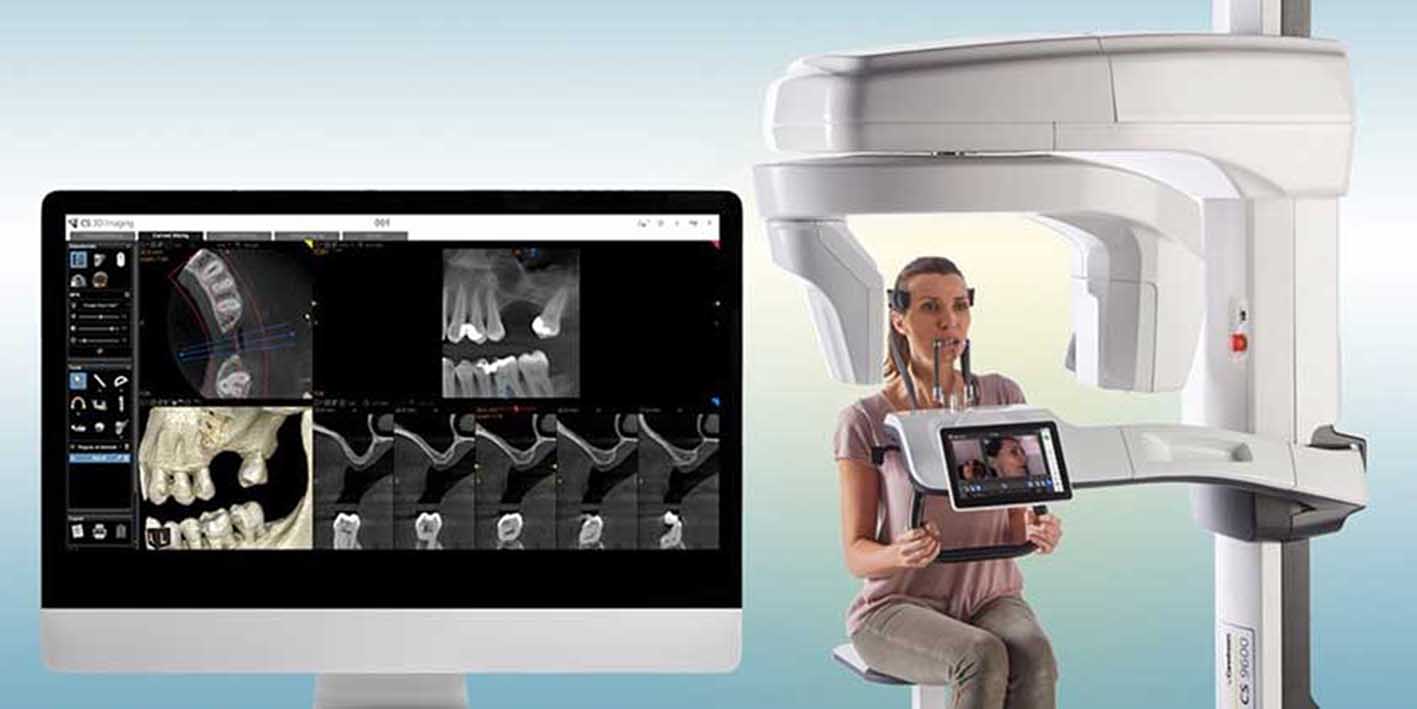
The CS 9600 from Carestream Dental is the next generation of CBCT systems, featuring never before achieved 2D and 3D imaging quality. It offers a multi-function imaging system blending 2D panoramic and cephalometric imaging, CBCT imaging, 3D facial scanning and 3D model scanning (some of these require optional upgrades). Designed to deliver more accurate scans on the first try, with consistent results over time, the system features AI-powered positioning which records each patient’s parameters, making follow-up imaging repeatable with a single touch.
In a world where dental technology is developing at pace, CBCT machines are transforming the way dentists work. Implementing one in your practice can help put you ahead of the curve in dental care.
 For more information on Carestream Dental visit www.carestreamdental.co.uk
For the latest news and updates, follow us on Facebook and Instagram @carestreamdental.uk

[i] The Society of Radiographers https://www.sor.org/learning-advice/professional-practice/diagnostic-imaging-pathways/dental-imaging
[ii] The Oral Health Foundation https://www.dentalhealth.org/x-rays
[iii] Shah N, Bansal N, Logani A. Recent advances in imaging technologies in dentistry. World J Radiol. 2014 Oct 28;6(10):794-807. doi: 10.4329/wjr.v6.i10.794. PMID: 25349663; PMCID: PMC4209425. [Accessed August 2023]
[iv] Mozzo P, Procacci C, Tacconi A, Martini PT, Andreis IA. A new volumetric CT machine for dental imaging based on the cone-beam technique: preliminary results. Eur Radiol. 1998;8(9):1558–64. 10.1007/s003300050586
[v] White S, Pharoah M. Chapter 13. Advanced Imaging Modalities. Oral Radiology: Principles and Interpretation. 5th ed. Mosby: St Louis, MO; 2004. pp. 245–264.
[vi] Venkatesh E, Elluru SV. Cone beam computed tomography: basics and applications in dentistry. J Istanb Univ Fac Dent. 2017 Dec 2;51(3 Suppl 1):S102-S121. doi: 10.17096/jiufd.00289. PMID: 29354314; PMCID: PMC5750833. [Accessed August 2023]
[vii] Almog DM, LaMar J, LaMar FR, LaMar F. Cone beam computerized tomography-based dental imaging for implant planning and surgical guidance, Part 1: single implant in the mandibular molar region. J Oral Implantol. 2006;32(2):77–81. 10.1563/789.1
[viii] Kapila S, Conley RS, Harrell WE Jr. The current status of cone beam computed tomography imaging in orthodontics. Dentomaxillofac Radiol. 2011. January;40(1):24–34. 10.1259/dmfr/12615645 [Accessed August 2023]
[ix] Cevidanes LH, Bailey LJ, Tucker GR Jr, Styner MA, Mol A, Phillips CL et al.. Superimposition of 3D cone-beam CT models of orthognathic surgery patients. Dentomaxillofac Radiol. 2005. November;34(6):369–75. 10.1259/dmfr/17102411
[x] Cotton TP, Geisler TM, Holden DT, Schwartz SA, Schindler WG. Endodontic applications of cone-beam volumetric tomography. J Endod. 2007. September;33(9):1121–32. 10.1016/j.joen.2007.06.011
[xi] Cotton TP, Geisler TM, Holden DT, Schwartz SA, Schindler WG. Endodontic applications of cone-beam volumetric tomography. J Endod. 2007. September;33(9):1121–32. 10.1016/j.joen.2007.06.011
[xii] Misch KA, Yi ES, Sarment DP. Accuracy of cone beam computed tomography for periodontal defect measurements. J Periodontol. 2006. July;77(7):1261–6. 10.1902/jop.2006.050367







