


Effective steps towards successful full arch implants
Featured Products Promotional FeaturesPosted by: Dental Design 16th November 2023
Dr Ferhan Ahmed BDS, MFDS, MBChB presents a case of full mouth rehabilitation using upper and lower implant-supported restorations.
I‘ve always been a big proponent for minimally invasive surgeries, and most importantly a step-wise approach to patient management. By this, I mean treating the most complex of cases with the simplest solution available. Where this gets more difficult, an invasive treatment is often necessary – but I always ask myself, does it have to be?
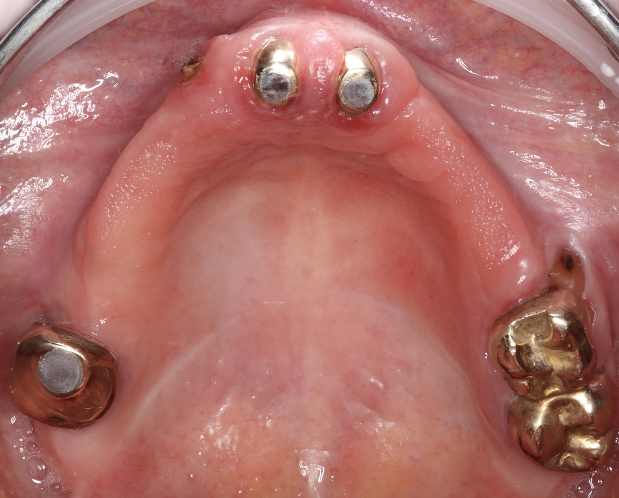
A 60-year-old female patient presented in a colleague’s practice with an upper denture, alongside already failing natural teeth, and a similar poor long-term prognosis for her teeth in the lower jaw. Her dentition had been treated and replaced over time, lacking consistency and an overarching plan. She was otherwise medically fit and healthy, and a non-smoker; her years as a GP had put her in good stead for great general health.
The patient expressed a desire for both upper and lower fixed solutions. She had already been through a consultation process elsewhere for this treatment but had not felt confident in them and their proposed plan.
All possible treatment options were discussed. We ruled out full removeable dentures, or utilising zygomatic implants alongside conventional implants. The patient’s desire for a fixed solution eliminated the possibility of new removable dentures, and we agreed that the latter left little room for potential future treatments.
We decided to use conventional implants with a palatal approach which was less invasive compared to utilising zygomatic implants. We discussed advantages and disadvantages of this approach, and the patient consented to proceed.
As part of a comprehensive examination and assessment for full arch surgery, medical history, periodontal status, occlusion and smile line were evalauated and no abnormalities were found. The emphasis on excellent oral health and nutrition in the recovery phase was discussed. As a healthy 60-year-old and GP herself, the patient needed little more than a recap.
In the days before the surgery, the patient was given suitable pre-operation medication, including analgesics and antibiotics, and then IV sedation and local anaesthetic on the day of the treatment.
A CBCT scan was used in pre-surgical planning to visualise the regional anatomy. The scan showed adequate bone height in zones 1 and 2 – premolar to premolar of the upper arch – however, the width of the ridge was very limited. Taking this into consideration, long tilted implants were used to maximise bone to implant contact, providing the best opportunity to achieve primary stability.
Digital software was used to assist in the planning of the surgical procedure, determining the ideal implant positioning, angulation and depth.
I opted to use Helix® Grand Morse® long implants from the Neodent® range. With various lengths available – 20mm, 22.5mm and 25mm – they provide optimal flexibility to maximise bone engagement and increase contact, which is essential for an immediate loading procedure.
During the surgery a flap was raised from the 7-7 region to allow good visualisation of all relevant landmarks, including the bulbosity of the maxillary sinus, floor of the nose, palatal vault and maxillary tuberosity.
The patient’s remaining teeth were extracted at the time of surgery. The bone was debrided and reduction of the ridge was performed to create a uniform, flat platform, ensuring enough restorative space for the bridge.
Regarding the palatal approach, it was important to enter in the premolar region from the palate, aiming buccally up towards the lateral nasal bone area. This bone was of high quality, therefore ideal for implants to engage and predictably provide stability. Drills involved in preparation of the osteotomy were the precision drill, followed by twist drill long 2.35 mm, then the final drill was 3.5 mm in diameter. Both tilted implants were the 4.0 x 18mm Helix® Grand Morse® range from Neodent® with a torque of 50Ncm plus on each. Angulation was corrected with 30 degree 3.5 mm MUA.
Two anterior implants – 4.0 x 10mm Helix® Grand Morse® – were placed in the lateral incisor region. The bone in zone 1 was sufficient for axial implants, and 50Ncm plus of torque was once again achieved. Good torque on all four implants allowed us to deliver fixed teeth in a day, for an immediate impact to the patient’s life.
The lower arch was also treated with immediately loaded implants and this was carried out uneventfully.
We follow up with our patient at regular intervals to assess the healing, as well as to ensure that the patient is following the post-operative care and dietary recommendations.
This case highlights the importance of approaching patient care in a step-wise manner, with the intention to minimise the invasiveness of treatment where possible. It also displays the need to look ahead for potential solutions; it is a clinician’s responsibility to plan for the future needs of our patients.
The palatal approach was a less invasive solution, in this case, allowing the patient to pursue zygomatic implants should she need them in the years to come.
My advice to other clinicians carrying out full arch surgeries would be to always consider alternative solutions. Beginning with the least invasive treatment option allows you to progress if the situation calls for it. This means working through implant surgeries that ascend in difficulty and invasive nature: for example, conventional implants, tilted implants, trans-sinus, pterygoid implants, and then zygomatic implant solutions.
This scale may also apply to a clinician’s potential training path. Don’t offer extra-maxillary implants or immediate full arch loading protocols until you are comfortable with conventional full arch implant treatment.
I run courses that help clinicians looking to further their skill set and learn full arch treatments, and how to manage more advanced cases. You can find out more about my unique and in-depth courses at www.drferhanahmed.com.


For more information, please visit www.neodent-uk.co/portal
Author Bio:

Dr Ferhan Ahmed BDS, MFDS, MBChB is a dental implant surgeon who is dual-qualified in dentistry and medicine. He qualified from the Univerity of Glasgow with a BDS in 2005, and from the University of Liverpool with an MBChB in 2012. His practice in dental implants involves conventional cases, complex bone grafting and extra maxillary implants. Dr Ahmed is also an educator within the field of dental implants and surgical dentistry.







