



Great first impressions make a lasting result
Featured Products Promotional FeaturesPosted by: Dental Design 8th February 2024
You don’t get a second chance to make a brilliant first impression. Whilst the sentiment is usually applied to those moving jobs or meeting new people, it still rings true for dentistry.
Impressions are essential throughout restorative care, providing the base for an accurate diagnosis and treatment planning with meticulous detail. The final result of a restoration or prosthesis will rely heavily on the accuracy of the initial impression, for a close-to-perfect fit.[i] With a fantastic impression, patient outcomes can be optimised.
As such, it’s important to understand how clinicians can make the most of every impression, for excellent results and a satisfied patient.
A matter of material
Impression taking dates back to the 18th century, and has since become a pivotal fixture in modern dentistry.[ii]The choice of material will be unique for each professional and treatment planning process. Understanding the advantages provided and the accommodations needed for the many options is key.
For example, polyether is a hydrophilic material that is suitable for humid surroundings, and provides excellent detail as its wettability allows dental gypsum to flow easily in the impression.[iii] It has very good elastic properties; modern iterations have avoided the difficulties of removal from a patient’s mouth when fully set and rigid, and without by-products from a setting reaction, it maintains good dimensional stability.[iv] However, clinicians must be aware that, due to its hydrophilic properties, it requires specific storage in environments of less than 50% humidity.[v]
On the other end of the spectrum, a hydrophobic material like condensation silicone is also widely used. It undergoes a polycondensation process that releases alcohol, contributing to the contraction of an impression.[vi] It can create accurate impressions when poured quickly after use, and has good elastic restoration after removal from the mouth, but its contraction means the impression may be less precise with the passing of time.[vii]
The potential solutions are not limited to these examples and there may not be a single best impression material for all treatment planning processes. When a clinician tailors their choice to their advantage, brilliant results can be created.
Digital impression
Today, physical impressions are not used in every aspect of modern dentistry, with advances to technological solutions supporting new workflows. Of these revolutionised options, intraoral scanners have emerged as an option.
Notably, their use has been reported to provide significantly more efficient and faster results than conventional methods – in some cases – whilst maintaining comparable accuracy in results.[viii] They may be especially useful in short-span scans, but some studies are sceptical about their use with completely edentulous patients[ix] and over longer distance spans.[x] Despite this, they may still be optimal for a number of applications, such as attaining impressions for the development of crowns and bridges.
Further on from the allure of an intraoral scanner’s convenience, there are concerns surrounding their accuracy when developing prostheses that come into contact with local soft tissue. A variety of studies agree that digital measurements at a subgingival marginal level are sometimes not clinically acceptable, making them inappropriate in these cases, especially where a crown’s margins are positioned deep (1.5-2mm) into the sulcus.[xi]
Power in the preparation
When preparing for the impression itself, soft tissue management could be the difference between restoration failure and success. If a prosthesis closely interacts with the gingiva, it is essential that the initial impression takes account of this – if not, the restoration may face a high chance of failure.[xii]
To effectively prepare the site for the impression, clinicians must appropriately displace local soft tissue, fluids and debris through gingival retraction. Traditional methods utilise a retraction cord to displace the soft tissue, however this could damage the gingiva, and when placed in an already inflamed or damaged sulcus, pre-existing problems may be exacerbated.[xiii]
This has prompted some clinicians to seek out effective alternatives. Pastes, foams and gels are popular, being atraumatic to the gingival tissue, without residue, and a simple, time-saving application. Cordless techniques put less pressure on the gingival tissue,[xiv] meaning a fantastic impression can be created without compromising the patient’s wellbeing.
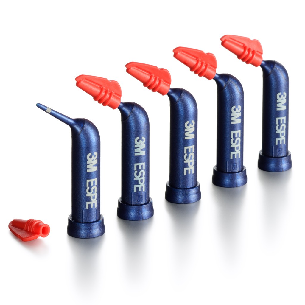
The ideal solution could be the 3M Astringent Retraction Paste, from 3M Oral Care, a preferred product for dental professionals*. The formula offers fast and precise gingival retraction, targeted through an extra-fine tip that fits right into the sulcus. Its long-lasting haemostatic effect makes it ideal on already damaged or lacerated soft tissue, for a comfortable and safe alternative to retraction cords.
Adapting the preparation process to each case’s needs is essential to creating an excellent impression that determines an even greater restoration. Today’s dental workflows offer the largest number of treatment solutions available to each clinician, but choosing the best tools throughout the process can help patients receive the greatest care possible.

For more information, call 08705 360 036 or visit www.go.3M.com/ocsdpr
3M is a trademark of the 3M Company.
*Dental Advisor 2023:
https://www.dentaladvisor.com/awards/2023-top-preferred-product-awards/#preferred-products
[i] Gupta, R., Brizuela, M., (2023). Dental Impression Materials. StatPearls [Internet] (Online) Available at: https://europepmc.org/article/NBK/nbk574496 [Accessed October 2023]
[ii] Goyal, G., Goyal, G., & Nagar, S. (2014). History of impressions, impression materials and impression techniques in complete dentures. Journal of Advanced Medical and Dental Sciences Research, 2(2).
[iii] Apinsathanon, P., Bhattarai, B. P., Suphangul, S., Wongsirichat, N., & Aimjirakul, N. (2021). Penetration and tensile strength of various impression materials of vinylsiloxanether, polyether, and polyvinylsiloxane impression materials. European Journal of Dentistry, 16(02), 339-345.
[iv] Apinsathanon, P., Bhattarai, B. P., Suphangul, S., Wongsirichat, N., & Aimjirakul, N. (2021). Penetration and tensile strength of various impression materials of vinylsiloxanether, polyether, and polyvinylsiloxane impression materials. European Journal of Dentistry, 16(02), 339-345.
[v] Gonçalves, F. S., Popoff, D. A. V., Castro, C. D. L., Silva, G. C., Magalhães, C. S., & Moreira, A. N. (2011). Dimensional stability of elastomeric impression materials: a critical review of the literature. European journal of prosthodontics and restorative dentistry, 19(4), 163.
[vi] Khan, S. A., Nezam, S., Singh, P., Kumari, N., & Singh, S. S. (2020). Comparison and evaluation of linear dimensional accuracy of three elastomeric impression materials at different time intervals using vision inspection system: an in vitro study. Journal of International Society of Preventive & Community Dentistry, 10(6), 736.
[vii] Naumovski, B., & Kapushevska, B. (2017). Dimensional stability and accuracy of silicone–based impression materials using different impression techniques–a literature review. Prilozi, 38(2), 131-138.
[viii] Afrashtehfar, K. I., Alnakeb, N. A., & Assery, M. K. (2022). Accuracy of intraoral scanners versus traditional impressions: A rapid umbrella review. Journal of Evidence-Based Dental Practice, 22(3), 101719.
[ix] D’Arienzo, L. F., & Borracchini, A. (2018). Comparison of the suitability of intra-oral scanning with conventional impression of edentulous maxilla in vivo. A preliminary study. Journal of Osseointegration, 10(4), 115-120.
[x] Kernen, F., Schlager, S., Alvarez, V. S., Mehrhof, J., Vach, K., Kohal, R., … & Flügge, T. (2022). Accuracy of intraoral scans: An in vivo study of different scanning devices. The Journal of Prosthetic Dentistry, 128(6), 1303-1309.
[xi] Casucci, A., Verniani, G., Habib, R., Ricci, N. M., Carboncini, C., & Ferrari, M. (2023). Accuracy of Four Intra-Oral Scanners in Subgingival Vertical Preparation: An In Vitro 3-Dimensional Comparative Analysis. Materials, 16(19), 6553.
[xii] Adnan, S., & Agwan, M. A. S. (2018). Gingival retraction techniques: a review. Dental update, 45(4), 284-297.
[xiii] Adnan, S., & Agwan, M. A. S. (2018). Gingival retraction techniques: a review. Dental update, 45(4), 284-297.
[xiv] Adnan, S., & Agwan, M. A. S. (2018). Gingival retraction techniques: a review. Dental update, 45(4), 284-297.







