



Let there be teeth!
Featured Products Promotional FeaturesPosted by: Dental Design 25th March 2024
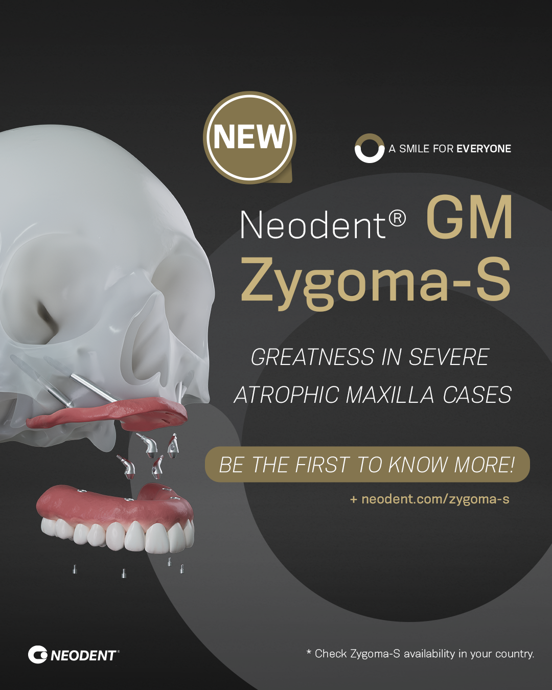
Dental implants offer a promising solution for edentulous patients with severe atrophic maxilla
Dental implants have revolutionised the field of dentistry by providing a reliable and long-term solution for patients with missing teeth. They offer numerous advantages over conventional dental treatments, such as dentures and bridges, including improved oral function, enhanced aesthetics, and increased patient satisfaction. However, placing dental implants in edentulous patients with severe atrophy of the maxilla presents several unique challenges that clinicians must navigate to achieve optimal results.[i]
Inadequate bone volume
 One of the major issues in treating edentulous patients with severe atrophic maxilla is the inadequate bone volume available for implant placement.[ii] The loss of teeth and the absence of dental roots cause the jaw bone to resorb over time,[iii] leading to a decrease in bone height and width. This bone loss compromises the stability and longevity of dental implants, as there may not be sufficient bone to anchor them securely. In such cases, bone augmentation and related procedures, such as sinus lift and ridge augmentation, are often necessary to create a suitable environment for implant placement.[iv]
Sinus lift is a surgical procedure performed to augment the maxillary sinus floor, which is frequently encountered in patients with atrophic maxilla.[v] Once the sinus membrane is lifted, bone grafting material is placed beneath it to create a thicker bone bed for implant placement. Ridge augmentation, on the other hand, involves adding bone graft material to the alveolar ridge to increase its width and height.[vi] Both procedures aim to enhance the quantity and quality of available bone, enabling successful implant placement in severe atrophic maxilla cases.
Another challenge in treating edentulous patients with severe atrophic maxilla is achieving adequate initial stability of dental implants. Reduced bone density and compromised bone quality can make it difficult to achieve sufficient implant stability during the initial placement. A potential solution to this challenge is the use of tilted implants and angulated abutments, which can engage areas of more substantial bone and provide improved distribution of forces. Utilising this type of implant and abutment can help overcome the limited available bone and maximise initial implant stability.[vii]
Soft tissue atrophy
The achievement and maintenance of an adequate soft tissue profile is also crucial in cases of severe atrophic maxilla.[viii] Edentulous patients often exhibit varying levels of soft tissue atrophy  and loss due to long-term denture use. Insufficient soft tissue volume can result in poor aesthetics and compromised peri-implant tissue health. Therefore, clinicians must evaluate and address the soft tissue deficiencies before implant placement. Techniques such as connective tissue grafting,[ix] guided bone regeneration,[x] and soft tissue augmentation[xi]can be employed to enhance soft tissue contour and optimise aesthetic outcomes.
Moreover, the treatment planning for edentulous patients with severe atrophy of the maxilla involves careful consideration of the prosthetic parameters. Occlusal forces exerted on dental implants can be significantly higher in these cases due to the lack of natural tooth support and increased bone density in the mandible.[xii]Therefore, providing adequate implant support and distribution of occlusal forces through the design of an implant-supported prosthesis is crucial. The prosthesis loading force is an important factor for dental implant survival. Computer-aided design and manufacturing (CAD/CAM) technology can be employed to fabricate a customised prosthetic framework that provides optimal support and stress distribution.[xiii]
Meeting edentulous patients’ expectations of shorter treatment times and immediate aesthetic and functional improvements present significant challenges for clinicians, especially in patients with anatomical deficiencies. The Neodent® GM Zygoma-S provides an immediate solution as an alternative for bone grafting procedures designed to deliver stability, treatment predictability, and precision, meeting patient expectations through its versatile design, and options tailored to anatomical efficiency and tissue relation. The protocol features unique drills that facilitate a more accurate osteotomy from the surgical beginning, bringing a maximum level of confidence to the surgery by precise initial drilling, preserving anatomical structures, and contributing to the accuracy in the implant positioning, regardless of the demanding indications.
Dental implants offer a promising solution for edentulous patients with a severe atrophic maxilla. However, these cases present unique challenges related to insufficient bone volume, inadequate initial implant stability, compromised soft tissue and prosthetic parameters. Yet, when addressed correctly, the dental clinician can overcome these difficulties and reach a successful outcome, enabling edentulous patients to smile once again with confidence.

For more details, please visit www.neodent-uk.co/portal
[i] Balan I, DI Girolamo M, Lauritano D, Carinci F. Treatment of severe atrophic maxilla with zygomatic implants: a case series. Oral Implantol (Rome). 2017 Nov 30;10(3):317-324. doi: 10.11138/orl/2017.10.3.317. PMID: 29285335; PMCID: PMC5735395. [Accessed December 2023]
[ii] Rues S, Schmitter M, Kappel S, Sonntag R, Kretzer JP, Nadorf J. Effect of bone quality and quantity on the primary stability of dental implants in a simulated bicortical placement. Clin Oral Investig. 2021 Mar;25(3):1265-1272. doi: 10.1007/s00784-020-03432-z. Epub 2020 Jul 10. PMID: 32651646; PMCID: PMC7878229. [Accessed December 2023]
[iii] Cemile Nurdan Ozturk, Can Ozturk, Mehmet Bozkurt, Halil Safak Uygur, Francis A. Papay, James E. Zins, Dentition, Bone Loss, and the Aging of the Mandible, Aesthetic Surgery Journal, Volume 33, Issue 7, September 2013, Pages 967–974, https://doi.org/10.1177/1090820X13503473 [Accessed December 2023]
[iv] Kumar, V.V., Ebenezer, S., Thor, A. (2021). Bone Augmentation Procedures in Implantology. In: Bonanthaya, K., Panneerselvam, E., Manuel, S., Kumar, V.V., Rai, A. (eds) Oral and Maxillofacial Surgery for the Clinician. Springer, Singapore. https://doi.org/10.1007/978-981-15-1346-6_19 [Accessed December 2023]
[v] Bathla SC, Fry RR, Majumdar K. Maxillary sinus augmentation. J Indian Soc Periodontol. 2018 Nov-Dec;22(6):468-473. doi: 10.4103/jisp.jisp_236_18. PMID: 30631223; PMCID: PMC6305100. [Accessed December 2023]
[vi] Kim YK, Ku JK. Ridge augmentation in implant dentistry. J Korean Assoc Oral Maxillofac Surg. 2020 Jun 30;46(3):211-217. doi: 10.5125/jkaoms.2020.46.3.211. PMID: 32606284; PMCID: PMC7338632. [Accessed December 2023]
[vii] Cavalli N, Barbaro B, Spasari D, Azzola F, Ciatti A, Francetti L. Tilted implants for full-arch rehabilitations in completely edentulous maxilla: a retrospective study. Int J Dent. 2012;2012:180379. doi: 10.1155/2012/180379. Epub 2012 Oct 23. PMID: 23133453; PMCID: PMC3486129. [Accessed December 2023]
[viii] Thoma DS, Gil A, Hämmerle CHF, Jung RE. Management and prevention of soft tissue complications in implant dentistry. Periodontol 2000. 2022 Feb;88(1):116-129. doi: 10.1111/prd.12415. PMID: 35103320; PMCID: PMC9306802. [Accessed December 2023]
[ix] Vatėnas I, Linkevičius T. The use of the connective tissue graft from the palate for vertical soft tissue augmentation during submerged dental implant placement: A case series. Clin Exp Dent Res. 2022 Oct;8(5):1103-1108. doi: 10.1002/cre2.626. Epub 2022 Jul 4. PMID: 35789972; PMCID: PMC9562572. [Accessed December 2023]
[x] Simoni Malushi E, Isufi R, Kadaifciu D. Guided Bone Regeneration Effects on Bone Quantity and Outcomes of Dental Implants in Patients With Insufficient Bone Support: A Single-Center Observational Study. Cureus. 2023 May 14;15(5):e38988. doi: 10.7759/cureus.38988. PMID: 37378176; PMCID: PMC10292071. [Accessed December 2023]
[xi] Hadzik J, Błaszczyszyn A, Gedrange T, Dominiak M. Soft-Tissue Augmentation around Dental Implants with a Connective Tissue Graft (CTG) and Xenogeneic Collagen Matrix (CMX)—5-Year Follow-Up. Journal of Clinical Medicine. 2023; 12(3):924. https://doi.org/10.3390/jcm12030924 [Accessed December 2023]
[xii] Kayumi, S., Takayama, Y., Yokoyama, A. et al. Effect of bite force in occlusal adjustment of dental implants on the distribution of occlusal pressure: comparison among three bite forces in occlusal adjustment. Int J Implant Dent 1, 14 (2015). https://doi.org/10.1186/s40729-015-0014-2 [Accessed Decmber 2023]
[xiii] Sangmyeong Tak, Gunwoo Noh, Yuwon Jeong, Hyeonjong Lee, Automated vector analysis to design implant-supported prostheses: A dental technique, The Journal of Prosthetic Dentistry, Volume 129, Issue 2, 2023, Pages 257-261, ISSN 0022-3913, https://doi.org/10.1016/j.prosdent.2021.05.017 [Accessed December 2023]







