The healing factor
Featured Products Promotional FeaturesPosted by: Dental Design 14th May 2020
The ability of the human body to heal itself is a remarkable thing. Much of medicine is an exercise in aiding or exploiting the body’s own healing and immunological processes. Surgery is thoroughly dependent on healing, without it – no matter how brilliant the technique – surgical intervention would be fruitless at best, if not outright harmful. Understanding, aiding and encouraging the body’s regenerative processes is therefore crucial to optimising interventions.
The oral mucosa is one of a rare set of tissues where virtually scar-free healing is possible in adult humans. Cuts that would leave disfiguring scarring on a patient’s skin can potentially heal to the point of near undetectability in the oral mucosa. The healing capabilities of the mucosa are of great benefit to surgery, both in aiding aesthetics and repair function. In the popular imagination, scars are often thought of as a mark of toughness, and their rougher texturing can give the impression that scarred areas are more resilient to future damage. In reality, this is not actually the case. Scarred skin will only ever achieve around 80% of its original strength at most, owing to a less optimal fibre structure that is significantly less elastic and more vulnerable to shear forces. Moreover, scarred skin will not regenerate functional features including pigmentation, sweat glands and hair follicles.[i] Though the mucosa has formidable regenerative capabilities, it can be inhibited by an absence of underlying healthy bone, thermal damage, and infection, or be unnecessarily degraded without careful surgical technique.[ii]
Saliva is highly beneficial to oral tissue healing, and may help healing elsewhere in the body (potentially explaining why animals evolved to instinctively lick their wounds). Saliva aids wound closure, increases cell turnover, encourages the release of growth factors and has numerous beneficial antimicrobial and anticariogenic properties.[iii], [iv] Over 99% of saliva is composed of water with the rest composed of proteins, ions and enzymes. The average person produces between 0.5 to 1.5 litres of saliva per day.[v] All patients benefit from making sure they are adequately hydrated by drinking water, as it will help maintain their saliva levels (not to mention benefitting kidney function and avoiding painful kidney stones). It is especially important when they are healing after oral surgery, at which time their oral hygiene is less than optimal.[vi], [vii]
In humans, the epidermal growth factor, basic fibroblast growth factor (bFGF), and insulin and insulin-like growth factor family have been detected in saliva, but their physiological role remains unclear. Recent studies, however, demonstrated beneficial effect of salivary bFGF on re-epithelization of mucosal epithelial defects.
Although saliva is a natural reservoir of growth factors, these can be supplemented following surgery with platelet derived growth factors. Activated platelet rich plasma (PRP) and platelet rich fibrin (PRF) products have been extensively employed to aid wound healing and tissue repair in different fields of medicine and surgery. These derivatives have different strengths; PRF provides a scaffold for tissue regeneration and healing, and has a consistent preparation protocol. PRP is enriched with PDGF-AB, which has an important role in wound healing and re-vascularisation. PDGF encourages bone cell replication and collagen production. Both PRP and PRF release significantly higher concentrations of growth factors than found in whole blood.[viii] Both platelet derivatives have been used to enhance the healing bone grafts and sinus augmentation procedures, as well as socket healing following tooth extraction and soft tissue injuries. Some research indicates PRP can aid osseointegration, though more research is needed to confirm this.[ix], [x]
Without proper case assessment aspects can be missed or overlooked, and the results for patients can be suboptimal, even disastrous on occasion. Particular care must be taken with patients who have failing implants, as previous surgery or infections can leave surrounding tissue scarred or damaged. Bone quantity and quality may have declined in the intervening time between procedures, potentially warranting grafting procedures.[xi], [xii] Dental implants placed in sites of previous implants that failed see a rise in reports of post-operative pain and a notably lower survival rate than initial implants (95.4% for initial implants, 77.4% for first reimplantation), which continues to decline with successive attempts (second reimplantation 72.7%, third 50%).[xiii] Of course, even first-time patients may have novel anatomical differences that could introduce complications if not accounted for. Understanding the case history, and having access to accurate and clear diagnostic imaging can do much to help optimise patient outcomes.
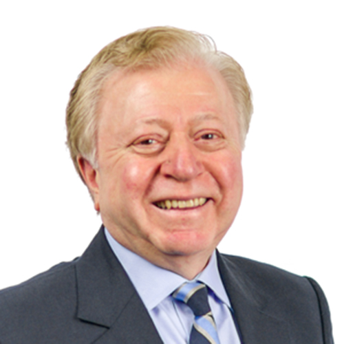
If you are dealing with a complex case, such as a failing implant requiring surgical intervention, consider referring your patient to the Centre for Oral-Maxillofacial and Dental Implant Reconstruction. Led by Professor Cemal Ucer – Specialist Oral Surgeon – the Centre offers a wide variety of advanced procedures, including nerve lateralisation and repositioning, major bone grafting, and rehabilitation of atrophic jaws with zygomatic ZAGA implants or 3D printed customised dental implants. With a wealth of experience – including in the use of platelet derived growth factor therapy – and state-of-the-art facilities you can be sure your patient will receive the very best of care.
Dental implants depend on the bone healing and bonding with the surface of the implant. Many factors can influence the osseointegration process, and some patients need additional surgery in order to meet the bone depth and quality required. Greater understanding of our healing processes and limitations facilitates improved treatment outcomes.
Please contact Professor Ucer at ice@ucer.uk or Mel Hay at mel@mdic.co
01612 371842
[i] desJardins-Park H., Mascharak S., Chinta M., Wan D., Longaker M. The spectrum of scarring in craniofacial wound repair. Frontiers in Physiology. 2019; 10: 332. https://www.ncbi.nlm.nih.gov/pmc/articles/PMC6450464/ February 7, 2020.
[ii] Politis C., Schoenaers J., Jacobs R., Agbaje J. Wound healing problems in the mouth. Frontiers in Physiology. 2016; 7: 507. https://www.ncbi.nlm.nih.gov/pmc/articles/PMC5089986/ February 7, 2020.
[iii] desJardins-Park H., Mascharak S., Chinta M., Wan D., Longaker M. The spectrum of scarring in craniofacial wound repair. Frontiers in Physiology. 2019; 10: 332. https://www.ncbi.nlm.nih.gov/pmc/articles/PMC6450464/ February 7, 2020.
[iv] Stookey G. The effect of saliva on dental caries. The Journal of the American Dental Association. 2008; 139(Suppl. 2): 11-17. https://jada.ada.org/article/S0002-8177(14)63877-0/fulltext February 7, 2020.
[v] Roblegg E., Coughran A., Sirjani D. Saliva: an all-rounder of our body. European Journal of Pharmaceutics and Biopharmaceutics. 2019; 142: 133-141. https://doi.org/10.1016/j.ejpb.2019.06.016 February 7, 2020.
[vi] Popkins B., D’Anci K., Rosenberg I. Water, hydration and health. Nutritional Review. 2010; 68(8): 439-458. https://www.ncbi.nlm.nih.gov/pmc/articles/PMC2908954/ February 7, 2020.
[vii] Roblegg E., Coughran A., Sirjani D. Saliva: an all-rounder of our body. European Journal of Pharmaceutics and Biopharmaceutics. 2019; 142: 133-141. https://doi.org/10.1016/j.ejpb.2019.06.016 February 7, 2020.
[viii] Arora S., Kotwal U., Dogra M., Doda V. Growth factor variation in two types of autologous platelet biomaterials: PRP versus PRF. Indian Journal of Hematology & Blood Transfusion. 2017; 33(2): 288-292. https://www.ncbi.nlm.nih.gov/pmc/articles/PMC5442062/ February 7, 2020.
[ix] Pirpir C., Yilmaz O., Candirli C., Balaban E. Evaluation of effectiveness of concentrated growth factor on osseointegration. International Journal of Implant Dentistry. 2017; 3: 7. https://journalimplantdent.springeropen.com/articles/10.1186/s40729-017-0069-3 February 7, 2020.
[x] Thalib B., Machmud E., Dharmautama M., Surya E., Hasyim A., Hasyim R. Differences of post-placement bone implant contact (BIC) value of dental implant coated and not coated with platelet rich plasma (PRP). Global Journal of Health Sciences. 2018; 10(2). https://doi.org/10.5539/gjhs.v10n2p11 February 7, 2020.
[xi] Sirajuddin S., Kripal K., Kumuda M., Chungkham S., Rafiuddin S., Chandrashekar B. Iatrogenic damage to the periodontium caused by implants and implant treatment procedures. Open Dentistry Journal. 2015; 9: 200-202. https://www.ncbi.nlm.nih.gov/pmc/articles/PMC4541314/ February 7, 2020.
[xii] Frizzera F., de Oliveira G., Shibli J., de Moraes K., Marcantonio E., Marcantonio Jr E. Treatment of peri-implant soft tissue defects: a narrative review. Brazilian Oral Research. 2019; 33(Suppl. 1). http://dx.doi.org/10.1590/1807-3107bor-2019.vol33.0073 February 7, 2020.
[xiii] Agari K., Le B. Successive reimplantation of dental implants into sites of previous failure. Journal of Oral and Maxillofacial Surgery. 2019. https://doi.org/10.1016/j.joms.2019.10.001 February 7, 2020.