


Prosthetically-driven, multidisciplinary dentistry
Featured Products Promotional FeaturesPosted by: Dental Design 20th September 2022
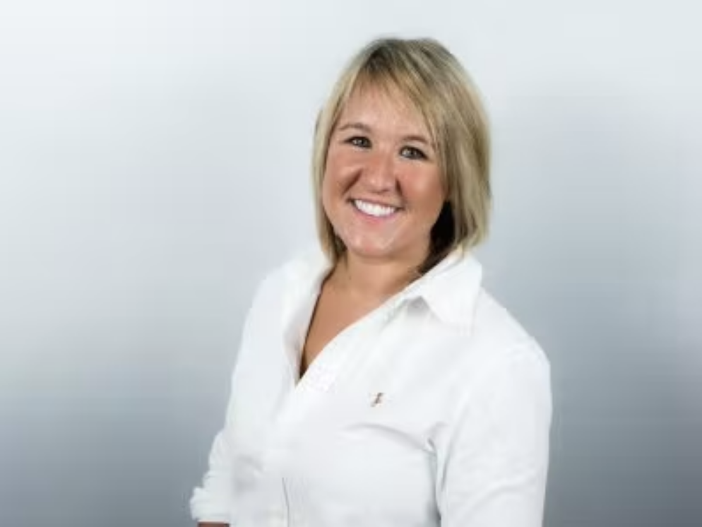
Dr Chloe Harrington-Taylor, a member of the highly talented team at Lodge Dental in South Wales, presents a comprehensive case that required a multidisciplinary, prosthetically-driven approach for the best results.
A 30-year-old female patient presented to Lodge Dental with a provisional bridge restoring a missing UL2, looking to improve her smile. She had a public-facing job and so sought a whiter, brighter and straighter smile to give her confidence in her role. She also expressed an interest in treatment that would disrupt or impact her job as little as possible too – requesting discreet and removable braces instead of fixed where possible.
Assessment
She was a regular attender every 6 months with the dentist/dental hygienist. Her medical and dental history was clear, she was a non-smoker and maintained an excellent standard of oral hygiene.
The initial smile consultation was completed including a comprehensive examination, which revealed the following:
- Class I molars on LHS
- Class II molars on RHS
- Class I canine on LHS
- Class II canines on RHS
- Class I incisor relationship
- 5mm overjet
- 30% overbite
- Missing tooth UL2
A facially-driven approach was taken when designing the treatment plan, which was created using the Invisalign Clin-Check software. This provided a visual representation of the potential outcome and ensured the patient was fully informed about the treatment process.
Once all possible treatment options were explained in detail, the patient chose to proceed with removable orthodontics (Invisalign), teeth whitening and a dental implant to replace the missing UL2. This procedure was explored in detail, with discussion of the potential risks, limitations and benefits of each stage of treatment. It was decided that the molar relationship would be accepted as the patient’s existing functional occlusion did not need to be altered. To keep the alignment process predictable, an aesthetic alignment of upper 5-5 was indicated.
All the necessary clinical photographs and digital impressions (using an iTero intraoral scanner) were taken and sent to the Invisalign for fabrication of the aligners. It was agreed to include a tooth in the aligners in the pontic space for the duration of orthodontic treatment so as not to impact the patient’s smile aesthetics and confidence for the time it would take to align the teeth. She was given careful instructions to wear the aligners for 22 hours a day, alongside oral hygiene support.
Alignment
The temporary bridge was removed and aligners were fitted at the first treatment appointment. The patient worked through the 14-aligner sequence, changing the aligner every 7 days as planned. Progressive interproximal reduction (IPR) was performed, and some attachments were placed to encourage the desired upper and lower alignment, with some minor rotation movements. It was important to remain mindful about maintaining appropriate space at the UL2 site for the later planned implant. The Bolton analysis was used to cross reference to the UR2 to deliver the best results and maintain symmetry. The orthodontic part of treatment concluded with a 2.1mm overjet and 30% overbite.
Throughout alignment treatment, virtual monitoring was utilised, which is typical for our orthodontic patients. The Virtual Care software allows the clinician to stay in better control of treatment without requiring the patient to visit the practice more regularly than absolutely necessary – it’s more efficient and saves time for both clinician and patient.
Implant treatment
Once the teeth were almost completely aligned, attention turned to implant placement. A CBCT scan was taken to assess bone volume, confirmation suitability of treatment and establish whether any augmentation would be needed.
As the patient had a medium smile line, aesthetics were an important consideration for this part of treatment, so a vertical relieving incision was made as a proven papilla-sparing flap design. A Southern Co-axis bone level implant was chosen in this case to allow for prosthetically-driven implant positioning and ideal restorative aesthetics.
In order to place an implant in the anterior maxilla you traditionally have to choose between a surgically-driven or prosthetically-driven implant position. Modern innovations such as angled screw correction have decreased our reliance on cement retained restorations and reduced the risk of associated peri-implant cementitis, but even angled screw correction has its potential downsides. Angled screw connection at implant level – which is possible with the co-axis implant design – allows correction of the implant angle and avoids the need for a cement-retained restoration or angled screw.
A synthetic bone graft with beta-tricalcium phosphate was performed alongside a connective tissue graft harvested from the palate.
The last seven finishing aligners were provided for the patient to wear while the implant was integrating. This ensured that everything was positioned perfectly as it healed.
Tooth whitening was performed towards the end of the refinement aligners, using Boutique Whitening for 2 weeks at night, placing the gel into the aligners – the patient’s smile went from A3 to B1.
The UL2 was restored at 12 weeks post-placement. Another digital scan was taken using the iTero scanner for this, before designing, fabricating and placing a screw-retained layered zirconia crown on a titanium base.
The importance of retention was emphasised with the patient, who will be wearing Invisalign retainers full time for 9 months and then at night lifelong.
Discussion
The patient development some mucosal scarring where the vertical relieving incision was made when raising the flap. If the patient had a high smile line, then de-epithelisation of the tissue could have helped to accelerate healing. However, her medium smile line meant this was not necessary – it will heal and appearance improve in time as it is. It is important to consider these aspects when treating younger patients whose dental implant may not last a lifetime.
This and any other potential complications must be address in the treatment planning stage – which is the most important aspect of any case, especially when prosthetically-driven results are the goal. All the pre-diagnostic planning from the wax-up to the surgical guides should correlate with the CT scan for success.

For more information about Dr Harrington-Taylor or Lodge Dental, or to refer a patient, please visit www.lodgedental.co.uk
Author:
Chloe graduated from the University of Bristol in 2013, where she received a distinction in restorative dentistry and was awarded the prestigious British Society for Restorative Dentistry Prize. Chloe has since achieved extensive postgraduate qualifications.
Chloe received membership to the Joint Dental Faculties with the Royal College of Surgeons and has a post graduate certificate in Aesthetic and Restorative Dentistry. As well as a Masters with Distinction from the University of Bristol for Dental Implantology alongside a Diploma in Clear Aligner Therapy.
Chloe’s clinical practice is limited to the provision of Invisalign orthodontic treatment and Implant prosthodontics. She has a comprehensive cosmetic and restorative portfolio.
She is a fully certified Invisalign Platinum provider and has a wealth of experience treating a full range of complex cases with Invisalign aligners.
Chloe takes referrals for Invisalign treatment and all aspects of implant dentistry working as part of a multidisciplinary approach to provide a full range of implant based restorative solutions, from single units to full arch fixed and removable implant supported dentures.







