


Double-arch implant reconstruction using Neodent GM Helix implants
Featured Products Promotional FeaturesPosted by: Dental Design 19th January 2023
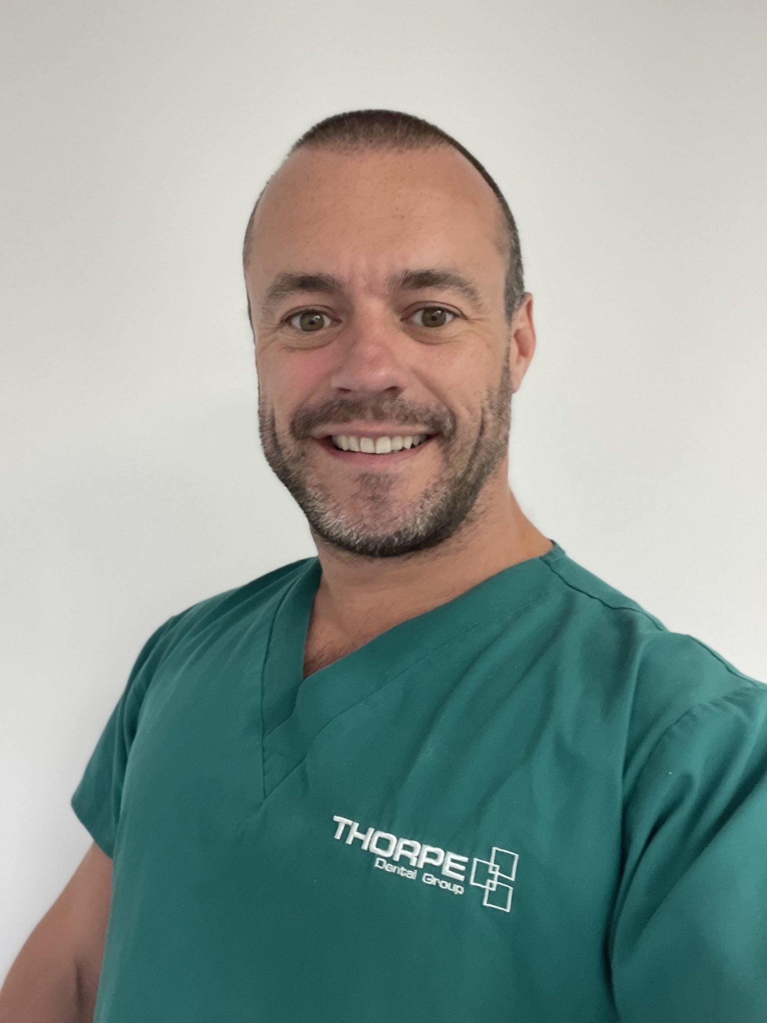
Dr Kris Leeson presents an implant resolution to a failing dentition.
This 61-year-old male patient first presented on 7th July 2021 to Thorpe Dental Implant Centre, having referring himself. He had expressed concern about his smile appearance and many missing teeth. He had a consultation in a practice in London who had advised that he could keep most of his remaining teeth and place implants in the missing spaces.
His main concerns in seeking treatment were: –
- Missing multiple teeth
- Felt embarrassed about his appearance and hid his smile
- Limits on the types of food he could eat
- Tired of feeling this way and wanted to change his life
Upon examination, the patient’s medical history was impeccable with no known allergies. However, his dental history indicated a significant amount of historical dental treatments and numerous extractions with no current prosthesis to restore his edentulous spaces. Due to this he had many over erupted teeth making a conformative approach almost impossible. He had a very deep traumatic overbite in several areas. His periodontal condition was poor with 90% root exposure UR6.
His lower dentition showed signs of severe wear. OPG revealed 100% bone loss UR6, retained root UL1. The retained root LL4 had associated apical pathology.
From initial examination it was quickly decided that he had a terminal dentition and all the remaining teeth would be better removed.
Treatment options
Following the assessment, a CT was taken, and treatment aims discussed. They were:
- To re-establish lower face height within normal and acceptable limits
- Improve function
- Eliminate infection
- Improve appearance and confidence
- Provide a fixed prosthetic result.
Following the clinical and initial radiographic assessment, it became clear that this case would require an upper and lower complete approach.
There were a number of possible treatment options open to us, as follows:
- Removal of hopeless teeth and restoration with a removable prosthesis
- Removal of hopeless teeth, placement of implants and restoration with a removable implant-supported / retained prosthesis
- Removal of hopeless teeth, placement of implants and restoration with a fixed prosthesis using a delayed approach.
- Removal of hopeless teeth, placement of implants and restoration with a fixed prosthesis using an immediate approach with an immediate provisional.
Following discussion with the patient, it was established that he wished for a fixed restoration and did not want dentures.
Treatment planning
After my initial consultation the patient was seen by one of the dental therapists at the practice. A digital impression, photographs and OPG were taken. After further discussion he decided the fixed immediate option was his preferred choice. It was at this discussion that a CT scan and impressions were taken and a bite recorded. These were recorded and sent to Harewood Dental Lab for an articulated set up.
Due to the patient high smile line and significant bone loss it was decided an FP3 approach would be the option of choice. This would enable the lost bone and soft tissue to be replaced with a pink material mimicking the gingival tissues.
From the CT scan it was determined that adequate bone was available for implant placement. No bone was available below the sinuses but angling implants into the premaxilla would give an adequate anterior posterior spread. Adequate bone was also available to reduce the alveolar ridge and hide the transition line when the patient smiled.
The patient journey
After the initial referral and examination, the patient was welcomed by my orthodontic therapist, who took a series of photographs and an OPG.

Figure 1 – pre op radiograph
It was decided that he wanted to explore further the possibility of an immediate implant fixed solution. From here a CT scan of the upper and lower arches was taken and implants planned on Galaxis implant planning software.

Figure 2 – Implant position planning on Galaxis software

Figure 3 – patient on presentation right lateral view

Figure 4 – patient on presentation left lateral view

Figure 5 – patient on presentation anterior view

Figure 6 – pre treatment facial view
Having gathered all the necessary records, impressions were sent to the laboratory for a mock up and fabrication of the relevant surgical stents.
The patient had requested sedation so a full sedation assessment was completed and the appropriate consent forms signed for both sedation and the implant procedure.

Figure 7 – Implants placement

Figure 8 – Implant placement
The surgery was complex but uneventful. Nine Neodent GM Helix implants were placed. These implants offer great primary stability and a great range of multi-unit components are available. The surgical stages for placing this implant are simple and all implant widths accept the same implant-abutment connection which makes everything easier.
All surgery and restoration was completed by mid-afternoon and the patient left happy with a new set of provisional teeth.

Figure 9 – Post implant placement radiograph

Figure 10 – Provisional restorations in place
The provisional restorations were kept in place for 3 months to allow osseointegration. When they were removed, the soft tissue was pink and healthy with adequate keratinised tissue around the mini conical abutments.

Figure 11a – Provisionals removed after three months

Figure 11b: Provisionals removed after three months
After 3 months, a duplicate of the provisionals were used to record the occlusion and a verification jig used to take the final silicone impression.

Figure 12 – silicone impression
The following appointments included a wax try in and metal try in followed by the final fit of the restorations. This case was restored with a titanium bar and acrylic wrap. A soft bite splint was also made to help protect the prosthesis from fracture whilst the patient was asleep.

Figure 13 – titanium bar with acrylic wrap

Figure 14 – Final restoration

Figure 15 – End result radiograph
Maintaining success
To maintain these appliances and the dental implants, the patient was advised to see a dental hygienist on a regular basis. For professional and at-home care I recommended:
- 3/12 dental hygiene appointments
- Oral hygiene using Idontix X-Floss and a Waterpik Water Flosser
- Night wear of a lower soft bite splint
- Occasionally, the bridges are to be removed and steam cleaned, with removal of any calculus deposits. The regularity of this is on a case-by-case basis.
This patient’s situation presented a number of interesting challenges, however, by using CHROME GuidedSmile we were able to achieve a very good outcome and sent away a smiling patient!
Overall, the patient had:
- Over-eruption of teeth
- Periodontal disease
- Apical pathology
- Traumatic overbite
- Lack of posterior support
- Loss of vertical stop
- Decrease in lower face height.
- Poor aesthetics
- Limited function
For more information about Neodent and the solutions available, please visit the website
Kris Leeson BChD MJDF MSc (Implant dentistry) PGCert (Health Care Ed) graduated from Leeds University in 2005. An award-winning dentist, he is also a member of the Faculty of Dental Surgery of the Royal College of Surgeons of England and has achieved a Master’s in Implant Dentistry, from Warwick University. He offers a full range of dental implant solutions, from the simple to the very complex. Kris has been involved in digital dentistry for ten years using digital solutions for nearly all his dentistry.







