


Is CBCT a good investment for your practice?
Featured Products Promotional FeaturesPosted by: Dental Design 23rd December 2023
Within a profession which is constantly developing thanks to the continuous integration of new technology, making wise investments is an excellent way to stay ahead of the competition. However, sometimes, this can be easier said than done. With so many options available on the market, it may be difficult to assess which options are most appropriate for your unique dental practice. The right investment may be the key to boosting diagnostics and treatment success, and helping more patients say ‘yes’ to treatment.
Benefits of CBCT
While cone-beam computed-tomography (CBCT) scanners are not an essential piece of equipment for all dental practices, for many they are ideal for improving diagnostic capabilities and treatment planning, and for carrying out regular treatment reviews. CBCT scanners produce highly accurate images which enable clinicians to clearly visualise the clinical situation, and make accurate assessments – proving very useful across a range of clinical scenarios including dental implants, orthodontics, and endodontics. It is important to take into account the risks of radiation in every case, and CBCT should only be used if the benefits outweigh any potential risks.[i] The recommendation in any case is to use the smallest possible field of view, and the lowest radiation exposure to reduce any risks to your patients. Cases should also always be selected carefully, to ensure that imaging is justified and benefits the patient.i
Dental implant practices
In practices which regularly provide dental implant treatment, it can be very helpful to have a CBCT scanner, as imaging is usually required throughout the treatment process. CBCT may be used to evaluate the linear measurements of the ridge height, width, and bone quality, assess the surface topography of the alveolar ridge, as well as illustrate the relevant vital anatomical structures, and recognise any incidental pathology (such as airway asymmetry, endodontic lesions, and temporomandibular joint disorder).[ii]
Their accuracy and image clarity can be ideal for diagnosis, allowing clinicians to firmly conclude whether dental implant treatment is appropriate. It can also improve treatment planning, when used alongside relevant software, high-quality imaging ensures accuracy. This can also improve treatment acceptance as patients are able to clearly understand their treatment plan, allowing the clinician to deliver a clear explanation.
Use in endodontic treatment
CBCT imaging is particularly useful in the diagnostic stages of endodontic treatment. Scans are able to improve diagnostic accuracy, and increase confidence in diagnosis, decision making, and treatment planning. CBCT Imaging may be used to asses for dental trauma, root resorption, vertical root fractures, and root canal anatomy. This allows clinicians to provide a more accurate diagnosis, and develop the most appropriate treatment plan for each individual case.[iii]
Application in orthodontic cases
CBCT imaging can make orthodontic treatment more predictable, allowing you to treatment plan with accuracy, and enabling patients to have a clear idea of their clinical situation, and of how their treatment will likely progress.  This, in turn, helps to manage patient expectations, increase acceptance, and boost patient satisfaction. This type of imaging, when used in orthodontics, is commonly indicated to assess anomalies of dental positions, like impacted or ectopic teeth for example, as it allows the visualisation of impacted teeth in detail, offering a three-dimensional view of the situation.i
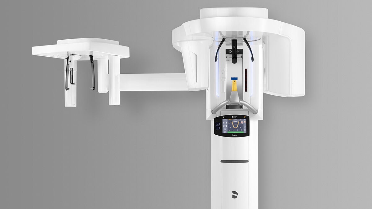
Having a CBCT scanner in the practice will reduce the number of referrals needed, and therefore the number of appointments patients are required to attend, and accessing imaging quickly will mean that treatment planning can proceed as soon as possible. When you choose the Axeos 2D/3D specialist system, available through Clark Dental, you can experience the best of 2D and 3D imaging, all in one device. The Axeos offers 4 volume sizes, from 5cm x 5.5cm to 17cm x 13cm, giving you more flexibility for a range of applications.
Investing in the most appropriate equipment for your unique dental practice will inevitably improve treatment workflows and outcomes, and lead to the increased satisfaction of your patients. While it is important to consider the effects that radiation may have on patients, weighing up the benefits and risks, in cases where it is appropriate, CBCT imaging serves as a useful diagnostic tool across a range of clinical scenarios.

For more information call Clark Dental on 01268 733 146, email info@clarkdental.co.uk or visit
STUART CLARK
MANAGING DIRECTOR

Having grown up within dentistry, Stuart has developed an unparalleled understanding of the industry. He is a technical specialist in digital imaging, pulling on his extensive experience as a Technical Director for Schick Technologies. Stuart is also an expert auto-CAD surgery designer and dental equipment engineer.
[i] Abdelkarim, Ahmad. “Cone-beam computed tomography in orthodontics.” Dentistry journal 7.3 (2019): 89.
[ii] Rios, Hector F., Wenche S. Borgnakke, and Erika Benavides. “The use of cone‐beam computed tomography in management of patients requiring dental implants: an American Academy of Periodontology best evidence review.” Journal of periodontology 88.10 (2017): 946-959.
[iii] Patel, S., et al. “Cone beam computed tomography in Endodontics–a review of the literature.” International endodontic journal 52.8 (2019): 1138-1152.







