


Can you keep pace with today’s evolving workflow for zygomatic implants?
Featured Products Promotional FeaturesPosted by: Dental Design 25th January 2024
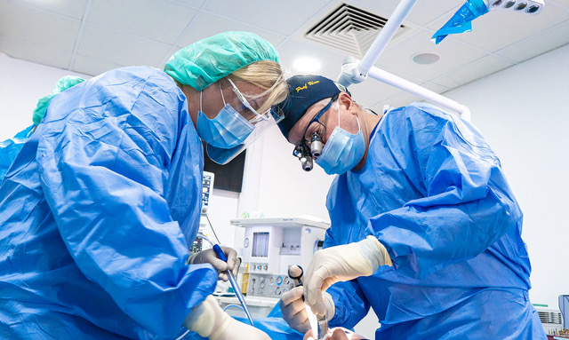
Zygomatic implant placement, often seen as a complex procedure for dental professionals, has evolved into a reliable solution for patients with an atrophic maxilla.
First introduced in 1998 by Professor Per Ingvar Brånemark, who is often referred to as the “Father of Dental Implantology”,[i] the treatment process has significantly improved over time, leading to higher success rates and fewer complications. Today, it is established as a predictable implant rehabilitation option in moderate to severely atrophic maxillary cases. Long-term survival rates have risen from 86% a decade ago[ii] to approximately 96.1% success after five years, according to recent studies.[iii] The success of zygomatic implants (ZIs) is expected to continue increasing with the recent introduction of the ZAGA-Straumann ZI, developed by Dr. Carlos Aparicio, founder of the ZAGA Centres.
The ZAGA philosophy emphasises anatomically guided ZI placement to preserve the integrity of the maxillary structures, including the maxillary sinus, whilst taking into account each patient’s unique maxillofacial anatomy. The combined use of the ZAGA Concept and the new ZAGA-Straumann implant designs has shown to result in less traumatic osteotomy, improved implant stability, better bone-to-implant contact, and enhanced bone sealing around the implant neck.[iv] Moreover, the rate of late complications, such as oral–sinus communication or soft tissue recession, has significantly decreased compared to the original technique.[v]
The advent of 3D digital technology has also played a crucial role in the improved success of ZIs. Today’s standard ZI procedure is markedly different from the first one performed over 25 years ago.
Into three dimensions
The planning stages of ZI placement are just as critical as the surgical procedure itself.
Today, 3D planning based on cone beam computed tomography (CBCT) is at the forefront of implant surgery. When used to inform a guided approach, it has been shown to increase the surgical accuracy and precision compared with free hand surgical techniques of the past.[vi]
With CBCT scans, clinicians can develop tailored treatment plans for each patient, taking into account the unique variations in facial structures, especially following atrophy in the maxilla.
 A ZI approach is distinctive in its own regard. By basing the implant in the namesake bone, clinicians enter new territories of facial surgery that open up the potential for unfavourable results. Whilst failure has been minimised, complications such as maxillary sinusitis may still arise at the hands of an inexperienced clinician or ill-informed treatment plan – when present alongside other issues, there may be a reduced chance of implant survival and/or a decreased quality of life.[vii]
One preferable approach to avoid rhinosinusitis complications is an extra-maxillary placement technique.[viii]This can help to avoid perforations of the sinus, which would produce the opportunity for infection. Accurately placed outcomes not only rely upon pre-operative 3D imaging, but digital planning and guiding software could be of use, too.[ix]
As part of the digital revolution in dentistry, computer-guided implant procedures look to establish themselves as a new normal in many workflows. This includes the use of a bone-supported guide, born out of digital preparation and 3D printing, which minimises the differentials of a planned and positioned implant.[x]
Ensuring minimal deviation between a planned approach and final outcome is essential. If the entry position or angle were to differ even minutely from the prospective design, a clinician could see a greater emphasised divergence at the apex. This issue is exacerbated in zygomatic procedures given the inherent length of the implant fixtures.[xi] The use of computer- and software-guided surgical approaches could therefore minimise the risk of intrusion into anatomical structures such as the maxillary sinus, minimising the development of sinusitis, and more closely replicating treatment plans.
Knowledge is power
Digital workflows do not make ZI placement any easier for the uninitiated, and the procedure must only be completed by those with the appropriate clinical training. Similarly to how treatment outcomes are only successful in the hands of a skilled clinician, 3D imaging from CBCT scanners and digital planning techniques are only beneficial when applied correctly.
Dental professionals should seek out education on the latest workflows and applications to ensure their patients with atrophic maxillae receive the highest quality of care. The training should be provided by experienced and trusted professionals, who have seen how workflows have developed and understand how to achieve optimal outcomes in modern dentistry.
Clinicians can begin their ZI journey with the hands-on course from Ucer Education based at ZAGA Centre Manchester which offers a comprehensive programme led by the acclaimed Specialist Oral Surgeon, Professor Cemal Ucer. With 25 years of teaching, research and clinical experience in implantology, the former ADI president ensures delegates understand the complete zygomatic workflow to implement the care into their practice. There is immense coverage on the use of modern CBCT imaging and 3D digital planning in the unique workflow, to help delegates plan effectively for their treatments.
Approaches to ZIs will only develop with time, but clinicians must embrace the capabilities of digital technology to further improve their treatment outcomes. With greater imaging and planning workflows, there is the opportunity to create safer and more successful solutions for those in need.
Please contact Professor Ucer at ucer@icedental.institute or Mel Hay at mel@mdic.co
01612 371842

Professor Cemal Ucer (BDS, MSc, PhD, Oral Surgeon, ITI Fellow)
Cemal Ucer first established an implant referral centre in 1995. He was awarded an MSc in Implantology at Manchester Dental Hospital following his research into guided bone regeneration and osteopromotion. He later gained a PhD for his clinical and laboratory studies into the factors affecting the success of implant treatment in iliac grafts and the investigation of the effect of skeletal bone density on implant survival. He has personally trained and mentored more than 1,000 dentists in implant dentistry as one of the main providers of implant education in the UK.
Cemal’s current clinical research interests include immediate implant placement, reconstructive bone surgery, nerve damage and the effect of bone density on the success of implant treatment. Academically, he has gained European recognition for his work on the development of a new framework for teaching and assessment of clinical competence in implantology. He is a co-author of the consensus paper produced by the Association for Dental Education in Europe (ADEE) following the first pan-European collaboration between EU universities to establish common training and assessment standards in dental implantology. He is an invited member of the working group convened by the FGDP (UK) and the General Dental Council (GDC) to update the Training Standards in Implant Dentistry (TSID) guidelines in 2012 and 2016.
Cemal is a Fellow of the Dental Trainers Faculty of the Royal College of Surgeons of Edinburgh (RCSEd) and a Fellow of the International Team for Implantology (ITI) and a member of Megagen’s MINTEC UK & I Board for education and clinical research. He is a member of the editorial board of JOMR (Journal of Oral & Maxillofacial Research) and the chair of the editorial advisory board of Implant Dentistry Today. Cemal is Professor and Clinical Lead of the MSc programme in Dental Implantology and a member of the Faculty of Examiners of the Royal College of Surgeons of Edinburgh’s Diploma in Implant Dentistry. He is a past president of The Association of Dental Implantology (ADI) (2011-2013).
Cemal has been appointed by FGDP (UK) to lead the working group to develop the “national standards in implant dentistry” which is due to be published later in 2018 following the completion of an external consultation process.
[i] Gaur, V., Doshi, A. G., & Palka, L. (2022). Zygomatic approach with single-piece implants: A technical note. National Journal of Maxillofacial Surgery, 13(1), 153.
[ii] Yates, J. M., Brook, I. M., Patel, R. R., Wragg, P. F., Atkins, S. A., El-Awa, A., … & Bolt, R. (2014). Treatment of the edentulous atrophic maxilla using zygomatic implants: Evaluation of survival rates over 5–10 years. International Journal of Oral and Maxillofacial Surgery, 43(2), 237-242.
[iii] Solà Pérez, A., Pastorino, D., Aparicio, C., Pegueroles Neyra, M., Khan, R. S., Wright, S., & Ucer, C. (2022). Success rates of zygomatic implants for the rehabilitation of severely atrophic maxilla: a systematic review. Dentistry Journal, 10(8), 151.
[iv] Aparicio, C., Polido, W. D., Chow, J., Davó, R., & Al-Nawas, B. (2022). Round and flat zygomatic implants: effectiveness after a 1-year follow-up non-interventional study. International Journal of Implant Dentistry, 8(1), 1-17.
[v] Aparicio, C., Polido, W. D., Chow, J., Davó, R., & Al-Nawas, B. (2022). Round and flat zygomatic implants: effectiveness after a 1-year follow-up non-interventional study. International Journal of Implant Dentistry, 8(1), 1-17.
[vi] Rinaldi, M., & Ganz, S. (2019). Computer-guided approach for placement of zygomatic implants: novel protocol and surgical guide. Compend Contin Educ Dent [Internet], 40(3).
[vii] Nocini, R., Panozzo, G., Trotolo, A., & Sacchetto, L. (2022). Maxillary sinusitis as a complication of zygomatic implants placement: a narrative review. Applied Sciences, 12(2), 789.
[viii] Solà Pérez, A., Pastorino, D., Aparicio, C., Pegueroles Neyra, M., Khan, R. S., Wright, S., & Ucer, C. (2022). Success rates of zygomatic implants for the rehabilitation of severely atrophic maxilla: a systematic review. Dentistry Journal, 10(8), 151.
[ix] Xing Gao, B., Iglesias-Velázquez, O., Gf Tresguerres, F., Rodríguez González Cortes, A., F Tresguerres, I., Ortega Aranegui, R., … & Torres, J. (2021). Accuracy of digital planning in zygomatic implants. International Journal of Implant Dentistry, 7(1), 1-8.
[x] Gallo, F., Zingari, F., Bolzoni, A., Barone, S., & Giudice, A. (2023). Accuracy of Zygomatic Implant Placement Using a Full Digital Planning and Custom-Made Bone-Supported Guide: A Retrospective Observational Cohort Study. Dentistry Journal, 11(5), 123.
[xi] Rinaldi, M., & Ganz, S. (2019). Computer-guided approach for placement of zygomatic implants: novel protocol and surgical guide. Compend Contin Educ Dent [Internet], 40(3).







