


Clinical success in the face of failure
Featured Products Promotional FeaturesPosted by: Dental Design 9th January 2024
Dental care can’t always be perfect. Sometimes, despite the best efforts of a clinician, a proposed treatment can face bumps in the road, or fail altogether. It’s therefore important to understand the risks to treatment success, as well as the actions to take when a case does face a ‘hiccup’.
In endodontics, this is no different. Ledge formation is a notable issue that occurs as a result of instrumental complications, and can create difficulties when continuing care – potentially even interfering with treatment success.
Endodontic jeopardy
A ledge is a deviation from the original pathway of a root canal, often occurring as a result of a clinical error during instrumentation.[i] This diversion can bring about a number of issues.
Successful endodontic treatment requires the thorough debridement and cleaning of the root canal system of any infected pulp tissue, which can then be shaped and prepared to be filled to prevent or minimise reinfection.[ii] A ledge can create complications within this clinical process by disrupting the guide of the canal. It may be difficult to achieve the adequate shaping of the canal to the ideal working length, resulting in incomplete disinfection of the structure, as well as insufficient filling.[iii]
It’s also possible that a ledge could initiate the creation of a perforation, if a clinician deviates from the root canal entirely.[iv] This may lead to infection of the periodontal ligament and alveolar bone;[v] both with catastrophic outcomes. If not properly detected and treated, the tooth may require extraction.
To ensure endodontic success can be achieved, and unfavourable treatment outcomes are avoided, clinicians must be able to identify the actions that lead to ledges to take appropriate pre-emptive measures.
Prevention is better than cure
Ledge formation is most likely to occur in the treatment of mandibular molar teeth.[vi] This is a result of their difficult accessibility and anatomical complexities when compared to anterior and premolar teeth. The structure of a mandibular molar is often prone to procedural aberrations, and when considering they often require the most involved endodontic treatment of any tooth, it’s no surprise that they yield the most ledges.[vii]
To prevent the creation of ledges, clinicians must approach endodontic care with an adequate pre-operative evaluation. Using radiographs to accurately identify the curvature, canal length and initial size of a site allow for a direct treatment plan, whilst giving the best opportunity to identify potential complications that may result in ledge formation.[viii]
Instrument choice is also key. Popular in modern endodontics, the use of flexible files results in a lower incidence of ledge formation when compared to conventional stainless steel K-type files.[ix] The development of nickel-titanium (NiTi) alloy instruments has been especially influential. They offer increased flexibility and result in fewer procedural errors, such as ledges, due to their superelasticity.[x] With the opportunity to bend the file prior to treatment, clinicians could experience a more controlled entry to and preparation of the root canal. When considering the complex, curved anatomy of mandibular molar teeth, the potential benefit is clear as day, but flexible instruments may also be ideal when treating patients with limited mouth openings.[xi]
Back to the wall
 It is likely impossible to complete an endodontic dental career without coming across a ledge. A clinician may have accidentally formed one, or could be treating a patient that has previously undergone care elsewhere, where the issue originated. Whatever the case, dental professionals must be prepared to approach treatment where it is present with an adequate solution.
If a clinician knows a ledge is present, they should first scout the space to locate the aberration, and identify the required approach to target the canal. Utilising an endodontic explorer may be most appropriate.[xii] Then, it may be necessary to bend a file toward the inside of the canal curvature, following the pre-determined angle that has been decided through the assessment.[xiii] With this guidance, the file will not divert into the ledge, and it can follow the canal to its apex.
This approach will be appropriate in most cases, as it is thought that ledge formation will mostly occur on the outer edge of the canal. Classically described, this is always the case, but beware rare exceptions.[xiv]
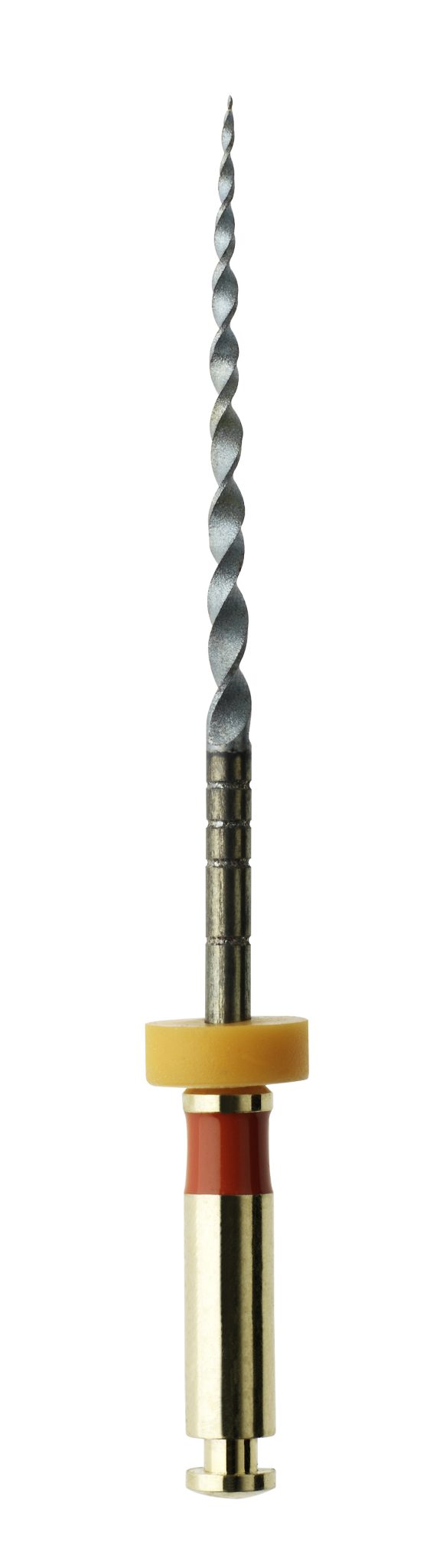
Prevention is always the priority, but clinical knowledge and excellent instruments can produce a brilliant outcome in the face of ledge formation. For confidence in their endodontic cases, clinicians should choose to equip the HyFlex™ EDM files from COLTENE. They provide unmatched flexibility and fracture resistance – up to 700% greater than a traditional NiTi file – and controlled memory properties allow the HyFlex™ EDM files to accurately follow the anatomy of the canal and reduce the risk of ledging, or potentially work around their presence.
Endodontic treatment is not free from adversity. With clinical expertise backed by the appropriate tools, dental professionals can still provide treatments that save patients a great deal of pain – even when the odds aren’t looking great.

For more information, info.uk@coltene.com and 0800 254 5115
COLTENE loyalty scheme https://rewards.coltene.com

[i] Cardinali, F., 2020. How to manage ledges using rotary files. Style Italiano Endodontics. (Online) Available at: http://endodontics.styleitaliano.org/how-to-manage-ledges-using-rotary-files/ [Accessed September 2023]
[ii] Tabassum, S., & Khan, F. R. (2016). Failure of endodontic treatment: The usual suspects. European journal of dentistry, 10(01), 144-147.
[iii] Jafarzadeh, H., & Abbott, P. V. (2007). Ledge formation: review of a great challenge in endodontics. Journal of endodontics, 33(10), 1155-1162.
[iv] Clauder, T. (2022). Repair of Pulp Chamber and Root Perforations. Endodontic Advances and Evidence‐Based Clinical Guidelines, 475-510.
[v] Nouroloyouni, A., Salem Milani, A., Etminan, A., Noorolouny, S., Tavakkol, E., Mikaieli Xiavi, H., & Ghoreishi Amin, N. (2023). Cone-Beam Computed Tomography Assessment of Quality of Endodontic Treatment and Prevalence of Procedural Errors in Mandibular Molars. International Journal of Clinical Practice, 2023.
[vi] Nouroloyouni, A., Salem Milani, A., Etminan, A., Noorolouny, S., Tavakkol, E., Mikaieli Xiavi, H., & Ghoreishi Amin, N. (2023). Cone-Beam Computed Tomography Assessment of Quality of Endodontic Treatment and Prevalence of Procedural Errors in Mandibular Molars. International Journal of Clinical Practice, 2023.
[vii] Berutti, E., Alovisi, M., Moccia, E., Carossa, M., De Caro, G., Roccuzzo, A., … & Scotti, N. (2022). Micro-computed tomographic evaluation of endodontic ledge position in relation to canal curvatures. BMC Oral Health, 22(1), 1-6.
[viii] Jafarzadeh, H., & Abbott, P. V. (2007). Ledge formation: review of a great challenge in endodontics. Journal of endodontics, 33(10), 1155-1162.
[ix] Jafarzadeh, H., & Abbott, P. V. (2007). Ledge formation: review of a great challenge in endodontics. Journal of endodontics, 33(10), 1155-1162.
[x] Tabassum, S., Zafar, K., & Umer, F. (2019). Nickel-titanium rotary file systems: What’s new?. European endodontic journal, 4(3), 111.
[xi] Tabassum, S., Zafar, K., & Umer, F. (2019). Nickel-titanium rotary file systems: What’s new?. European endodontic journal, 4(3), 111.
[xii] Terauchi, Y. (2008). Correction of ledged canals with ultrasonic tips. Dentistry today, 27(10), 140-145.
[xiii] Berutti, E., Alovisi, M., Moccia, E., Carossa, M., De Caro, G., Roccuzzo, A., … & Scotti, N. (2022). Micro-computed tomographic evaluation of endodontic ledge position in relation to canal curvatures. BMC Oral Health, 22(1), 1-6.
[xiv] Suciu, I., Ciocardel, M., Dimitriu, B., Ionescu, E., Chirila, M., Teodorescu, E., … & Amza, O. (2020). Microscopic study of endodontic ledges on extracted teeth. ROMANIAN BIOTECHNOLOGICAL LETTERS, 25(5), 2008-2017.







